Preview

Sechenov Medical Journal

Advanced search

The Sechenov Medical Journal is a scientific and practical peer-reviewed journal, the official publication of Sechenov University. 

The Journal has been published since 2010 with a frequency of 4 issues per year and is intended for the health professionals.


Sechenov Medical Journal publishes original articles, reviews, and clinical cases, covering a wide range of issues in biomedical sciences, fundamental and clinical medicine and concerned with important clinical and basic research in the field of:

  • cell biology,
  • pathological physiology,
  • internal diseases,
  • obstetrics and gynaecology,
  • oncology, surgery  
  • neurosurgery.

Publication time frames:

5 days - first decision (accept for review or reject the manuscript)
40 days - average duration of the review phase
99 days - from manuscript submission to publication (average)
27% - of all manuscripts submitted during the year were accepted for publication


The Title is included in the Russian Science Citation Index (RSCI) collection, based on the Russian Index of Science Citation(RISC) database and is in the Scopus database (since 2023) and in the "White List" of scientific journals

Sechenov Medical Journal was included in the K2 category of Higher Attestation Commission and equated to the K1 category considering indexing in Scopus.

Mass media state registration certificate PI № ФС77-78884 dated August 28, 2020, issued by the Federal Service for Supervision of Communications, Information Technology and Mass Media (Roskomnadzor).

Current issue

Vol 16, No 1 (2025)
View or download the full issue PDF (Russian)

CELL BIOLOGY, CYTOLOGY, HISTOLOGY

4-19 430
Abstract

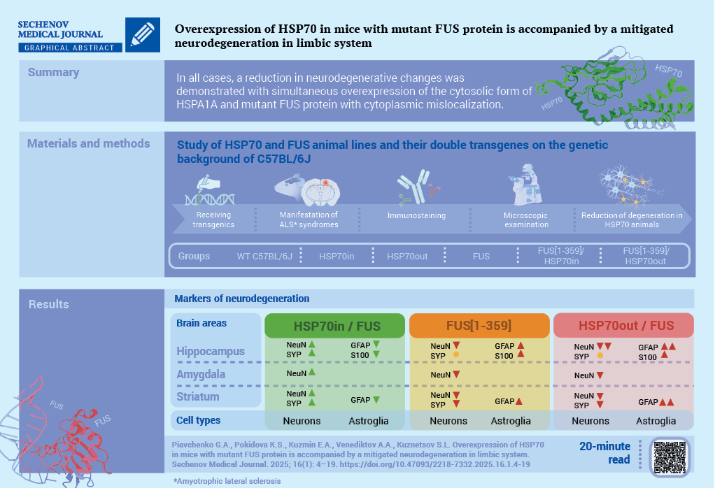
Aim. To study morphological and developmental changes in the structure of mice’s limbic system, which overexpress 70 kDa heat shock proteins (HSP70) and co-express mutant fused-in-sarcoma (FUS) protein with amyotrophic lateral sclerosis (ALS).

Materials and methods. The study was based on mice (n = 36; six for each group) of six lines either without FUS mislocalization: C57Bl/6 (wild-type); extracellular (HSP70out) or intracellular (HSP70in) overexpression of HSP70 family 1A protein; or with FUS mislocalization: transgenic mice with ALS-FUS (FUS[1-359]); and double transgenic animals (FUS[1-359]/HSP70out and FUS[1-359]/HSP70in). All FUS expressing mice were symptomatic with myasthenia up to limb paralysis in some animals. When the mice were 20 weeks old, they were killed by staining histological slides of their brain using hematoxylin and eosin, toluidine blue by Nissl, immunofluorescent antibodies for the neuronal nuclear marker (NeuN) of the caudoputamen, septal nuclei, and hippocampus, as well as glial fibrillar acid protein (GFAP), S100β protein, and synaptophysin for the hippocampus. The number of neurons and the number of cells with a positive reaction to antibodies were counted. The statistical processing included ANOVA and were compared with the results from the Tukey test for.

Results. Statistically significant differences were observed for the FUS-expressing groups compared to FUS-negative groups: (1) a reduction in the number of neurons and NeuN+-cells in the caudoputamen and amygdala, especially for FUS[1-359]/HSP70out group; (2) an increase in the number of hyperchromic neurons in the subiculum, cornu Ammonis (CA1), and dentate gyrus, with a significantly greater increase for FUS[1-359] and FUS[1-359]/HSP70out groups compared to FUS[1-359]/HSP70in group; (3) an increase in the number of GFAP+and S100β+-cells in the hippocampus, with the most pronounced change for the FUS[1-359] and FUS[1-359]/HSP70out groups compared to с FUS[1-359]/HSP70in group.

Conclusion. An overexpression of HSP70 family 1A protein and co-expression of mutant FUS protein in the cytoplasm is accompanied by mitigated neurodegeneration in the structure of the limbic system compared with the expression of mutant FUS protein alone.

PATHOLOGICAL PHYSIOLOGY

20-33 185
Abstract

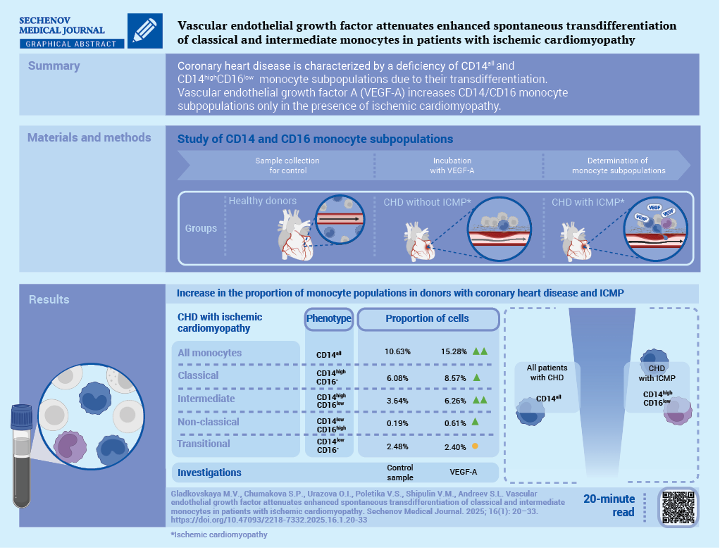
Aim. To evaluate the effect of vascular endothelial growth factor A (VEGF-A) on the subpopulation composition of monocytes in the blood mononuclear cell culture of patients with coronary heart disease (CHD), with and without ischemic cardiomyopathy (ICMP).

Materials and methods. A single-center, experimental in vitro study was conducted. The study included 22 patients with CHD: 11 with ICMP, 11 without ICMP, and 10 healthy donors. Blood mononuclei were isolated from venous blood by immunomagnetic separation for CD14 and CD34 antigens, then incubated with and without the addition of VEGF-A 50 ng/mL (control and stimulated samples). After 6 days, the total monocyte content, the proportion of classical CD14++CD16, intermediate CD14++CD16+, non-classical CD14+CD16++, and transitional CD14+CD16 monocytes were assessed using flow cytofluorimetry.

Results. In groups of patients with CHD and in those groups where the patients were considered relatively healthy, a decrease in the content of CD14++CD16+ in the control and stimulated samples was shown. Only in the CHD group with ICMP relative to the control sample, after VEGF-A stimulation, a statistically significant increase in all CD14+ was found: 10.63% (6.80; 17.64) vs. 15.28% (8.75; 27.99), p < 0.01, and their subpopulations: CD14++CD16−: 6.08% (1.76; 8.84) vs. 8.57% (3.51; 16.8), p < 0.05, CD14++CD16+: 3.64% (2.03; 8.59) vs. 6.26% (3.87; 10.3), p < 0.05. In the same group, a tendency towards an increase in CD14+CD16++ was noted after stimulation: 0.19% (0.18; 1.11) vs. 0.61% (0.37; 1.58), p = 0.062. No differences in the content of all monocytes and their subpopulations after VEGF-A stimulation were found in the CHD without ICMP group nor in the healthy group. The content of CD14+CD16 in all groups in the control and stimulated samples did not differ.

Conclusion. CHD is characterized by a deficiency of all CD14+ cells and intermediate monocytes due to their transdifferentiation. VEGF-A affects the subpopulation composition of monocytes in CHD only in the presence of ICMP by increasing the content of all CD14+ cells, and in their intermediate and classical forms without exceeding the indicators in healthy donors.

34-44 225
Abstract

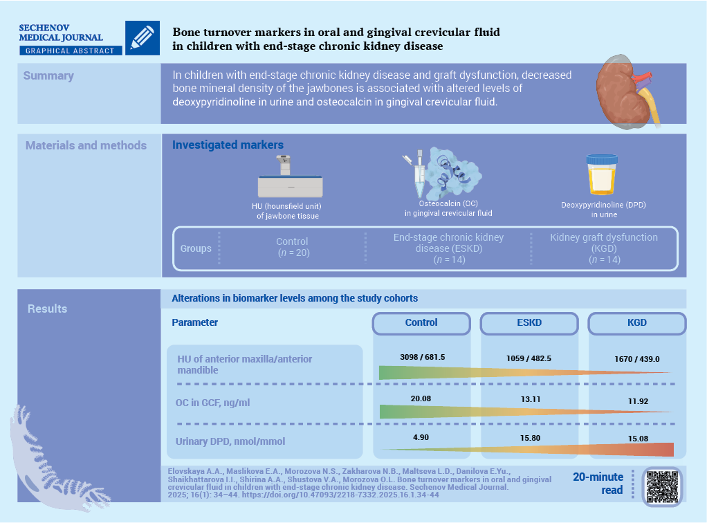
Objective. To study bone turnover markers in biological fluids (urine, blood serum, oral fluid (OF) and gingival crevicular fluid (GCF)) at the stage of planning an orthodontic strategy in children with end-stage chronic kidney disease (ESKD).

Materials and methods. Pilot, cross-sectional, multicenter study was conducted. A total of 48 children aged 7 to 17 years were examined and divided into three groups: 14 children with ESCKD, 14 children with renal transplant dysfunction (RTD), 20 almost healthy children. Bone turnover markers were assessed by changes in osteocalcin (OC) in the OF, GCF and blood serum, urinary deoxypyridinoline (DPD), levels of total, ionized calcium and phosphorus in blood and pH of OF. Bone tissue mineral density was assessed by cone-beam computerized tomography according to the C. Mish classification.

Results. All groups of children were comparable by gender and age. All patients had no significant mineral and bone disorders. Total and ionized calcium did not demonstrate statistically significant differences between the study groups. Serum phosphorus level was higher in ESCKD children compared to RTD children and control group. Urinary DPD, OC in GCF and OF pH were higher in children with CKD compared to healthy children. However, there were no statistically significant changes between the ESCKD group and the RTD group. In the posterior maxilla, the Hounsfield index was higher in the group with RTD compared to the ESCKD group (p < 0.01), and similar to the control group. In the anterior maxilla, as well as in the anterior and posterior mandibular regions, the Hounsfield index was higher in the control group than in the ESCKD and RTD groups.

Conclusion. The most prominent changes of bone turnover markers were found in children with ESCKD. Urinary DPD and OC in GCF were associated with the decrease in kidney function and jawbone mineral density.

INTERNAL MEDICINE

45-54 269
Abstract

Background. Type 2 diabetes mellitus (T2DM) poses a significant challenge to healthcare, with its prevalence escalating to epidemic proportions. The aging population, coupled with the increasing burden of T2DM, is exerting immense pressure on healthcare systems worldwide. Therefore, there is a critical need to design and validate innovative interventions to mitigate the effects of this disease. This randomised control trial aims to achieve remission in Indian patients aged 18 years and older diagnosed with T2DM through dietary and behavioural interventions.

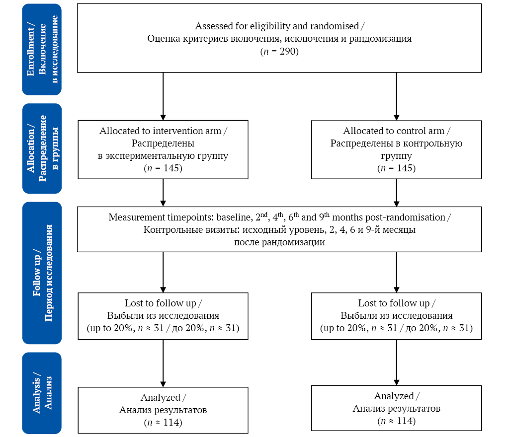
Materials and methods. A total of 290 participants with T2DM will be recruited from Indira Colony Urban Enclave, the field practice area of the Department of Community Medicine and School of Public Health at Post Graduate Institute of Medical Education and Research, Chandigarh. Participants will be equally allocated into two arms: intervention (n = 145) and control (n = 145). There will be five measurement timepoints: baseline, 2nd, 4th, 6th and 9th months postrandomisation. The intervention will implement a range of strategies to increase physical activity and promote dietary transitions through behaviour change among patients. The interventions will be designed ensuring a structured approach to behaviour change. Patients from the intervention arm will receive oral hypoglycaemic agents for the first six months of the trial. After this period, medication will be gradually tapered. Patients from the control arm will continue to receive standard care throughout the study. The primary outcome is the number of patients achieving remission of T2DM through behavioural and dietary interventions.

Conclusions. The novelty of this trial lies in its focus on community-based settings, unlike other studies that primarily target clinical or hospital-based environments to achieve clinical outcomes. The intervention integrates dietary and behavioural changes into the community’s cultural, socioeconomic, and dietary habits, making it practical and sustainable for patients to adopt and maintain the lifestyle changes needed for remission.

NEUROSURGERY

Announcements

2025-06-30

The social channels of the Sechenov Medical Journal were featured in a scientific article.

We are pleased to announce that the Sechenov Medical Journal's Telegram channel was mentioned in an article by M. A. Polshina and L. V. Vergunova, published in Scientific Editor and Publisher (https://doi.org/10.24069/SEP-24-24 ).

The authors cited our channel as a successful example of scientific journal social networking to increase citations of scientific publications.

The study confirms the effectiveness of scientific journals' activity on social networks:

- increases traffic to the journal's website

- increases the impact factor and altmetrics of publications

- makes content more accessible

- Promotes audience engagement

- increases citations of publications.


According to the authors' findings, the portrait of a subscriber to the Telegram channel of a scientific journal is as follows:

- an employee of a higher education institution or research institute.

- aged 26–45 years;

- holds a Candidate of Science degree,

- Hirsch index in RSCI is less than 9.

The most popular section is the 'Weekly review of world news'. Short posts of 2–3 paragraphs are the most effective format.


The editorial team at the Sechenov Medical Journal closely follows global trends in scientific communication, offering you only the most relevant and useful information.


Join our Telegram community (https://t.me/sechmedj ) and help us to make science more accessible!

More Announcements...


Creative Commons License
This work is licensed under a Creative Commons Attribution 4.0 License.