


INTERNAL MEDICINE
Lipoprotein (a) (Lp(a)) is a genetically determined risk factor for atherosclerosis, with extremely high levels associated with very high cardiovascular risk. Despite this, in routine clinical practice these patients often remain insufficiently identified.
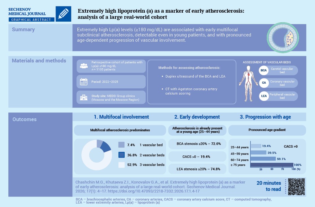
Aim. To characterize the prevalence and severity of subclinical atherosclerosis in various vascular beds in patients with Lp(a) ≥ 180 mg/dL, including younger age groups.
Materials and methods. We performed a retrospective analysis of a database comprising 101,078 outpatients, from which 1105 (1.09%) individuals with Lp(a) ≥ 180 mg/dL and available lipid profile data were selected; women accounted for 67.2%, and the mean age was 52.74 ± 14.59 years. The presence of atherosclerotic cardiovascular disease (ASCVD) was ascertained from medical records, and atherosclerosis was assessed by duplex ultrasound of the brachiocephalic arteries (BCA) and lower extremity arteries (LEA), as well as by the Agatston coronary artery calcium score (CACS). At least one vascular bed was evaluated in 69.8% of patients, and all three were evaluated in 6.2%.
Results. Most patients showed elevated levels of total cholesterol and low-density lipoprotein cholesterol. From the age of 25–44 years onward, a substantial proportion of patients already showed atherosclerotic involvement of the BCA and LEA with stenosis ≥20% and pronounced coronary calcification (CACS > 100 units), with a shift toward more severe lesions in older age groups. In the subgroup that underwent imaging of all three vascular beds, 60.0% of patients aged 25–44 years had two or three beds that were affected; the proportion of patients with involvement of all three vascular beds increased to 40.0% in those aged 45–59 years, 76.9% in those aged 60–74 years, and reached 100% in patients aged ≥ 75 years (p < 0.001). The prevalence of clinically documented ASCVD increased from 10.3% in the 25–44 age group to 67.1% in patients aged ≥ 75 years. Even among patients without documented ASCVD, atherosclerotic involvement of the BCA, LEA and coronary calcification was detected in 85.9%, 82.6% and 37.1% of cases, respectively.
Conclusion. Patients with Lp(a) ≥ 180 mg/dL are characterized by a high prevalence and early onset of subclinical, frequently multifocal atherosclerosis, with a pronounced age-related gradient of progression. These findings support the case for designating this cohort as a priority group for in-depth evaluation, risk re-stratification and more intensive preventive management.
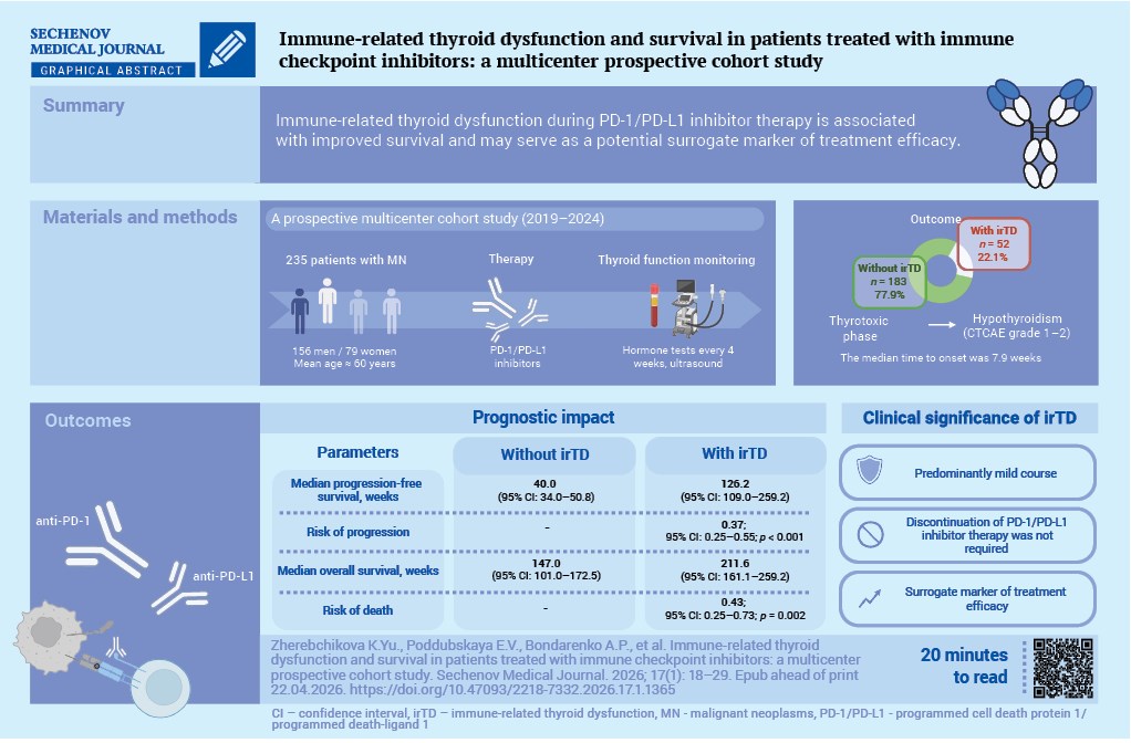
Aim. To assess the association between immune-related thyroid dysfunction (irTD) and overall survival or progressionfree survival in patients with malignant neoplasms receiving immune checkpoint inhibitor therapy.
Materials and methods. This multicenter prospective cohort study comprised 235 patients (156 men; mean age approximately 60 years) with histologically or cytologically confirmed malignancies of various localizations who were treated with programmed cell death protein 1/programmed death-ligand 1 (PD-1/PD-L1) inhibitors. Thyroid function was assessed before the treatment and every 4 weeks thereafter. irTD was diagnosed based on standardized biochemical and ultrasound criteria. Adverse events were graded according to CTCAE (Common Terminology Criteria for Adverse Events). Survival was analyzed using the Kaplan–Meier method, with comparisons performed using the log-rank test, and univariable Cox proportional hazards models, with hazard ratios (HRs) and 95% confidence intervals (CIs), were applied.
Results. irTD occurred in 52 patients (22.1%). In all cases, destructive thyroiditis with a transient thyrotoxic phase followed by hypothyroidism (CTCAE grade 1–2) was observed and did not require discontinuation of immune checkpoint inhibitors. The median time to irTD onset was 7.9 weeks. Patients with and without irTD were comparable in terms of sex, age, disease stage, previous cancer therapy, and type of PD-1/PD-L1 inhibitors. The development of irTD was associated with better progression-free survival (median 126.2 vs 40.0 weeks; HR 0.37; 95% CI: 0.25–0.55; p < 0.001) and better overall survival (211.6 vs 147.0 weeks; HR 0.43; 95% CI: 0.25–0.73; p = 0.002).
Conclusion. In this prospective multicenter cohort study, irTD occurred in approximately one-fifth of patients treated with PD-1/PD-L1 inhibitors, mainly during the first weeks of therapy, was generally mild, and was associated with improved survival. These findings suggest that irTD may be considered as a potential surrogate marker of treatment efficacy and support the need for regular monitoring of thyroid function during immunotherapy.
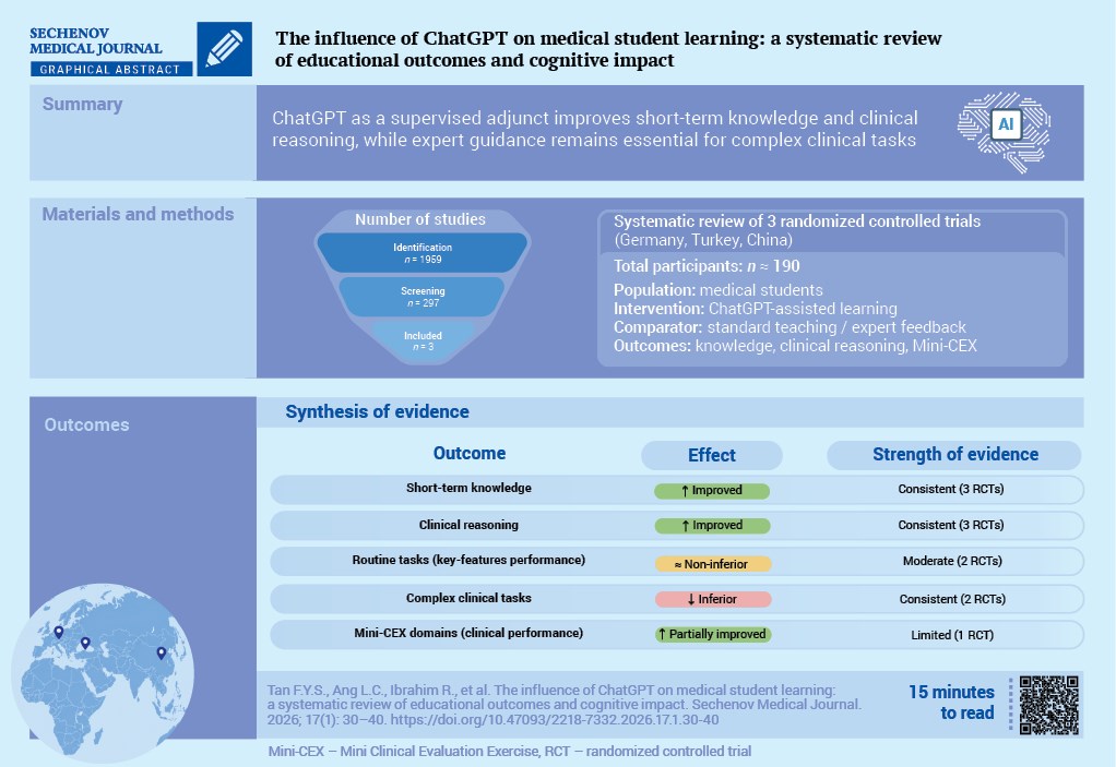
Aim. To evaluate whether the use of ChatGPT as a supplement to usual teaching improves medical students’ shortterm knowledge, clinical reasoning, and near-term performance.
Materials and methods. We systematically searched PubMed, Scopus, ScienceDirect, SpringerLink, and Web of Science on 25 June 2025, for studies involving medical students that evaluated ChatGPT as a supplement to teaching and reported objective educational outcomes. Two independent reviewers screened records, extracted data, and assessed the risk of bias. A narrative synthesis was then conducted due to the level of heterogeneity in interventions and outcome measures across the studies.
Results. Three randomized trials conducted in Germany, Turkey, and China met the inclusion criteria. ChatGPTsupported interventions improved or at least maintained short-term educational outcomes over the control groups for knowledge tests, clinical reasoning, and some of the Mini-Clinical Evaluation Exercise (Mini-CEX) domains.
Structured and immediate ChatGPT feedback improved Clinical Reasoning Indicator-History Taking Inventory scores after a simulated patient encounter, and ChatGPT-generated explanations were not inferior to expert feedback in overall key-features question performance but were less effective for more complex items, where expert feedback remained superior. Overall, the risk of bias was judged to be low to some concerns, with likely unblinded Mini-CEX assessment noted as a significant limitation.
Conclusion. ChatGPT used as a supervised adjunct to teaching showed value for short-term knowledge acquisition and clinical reasoning development.
NEUROSURGERY
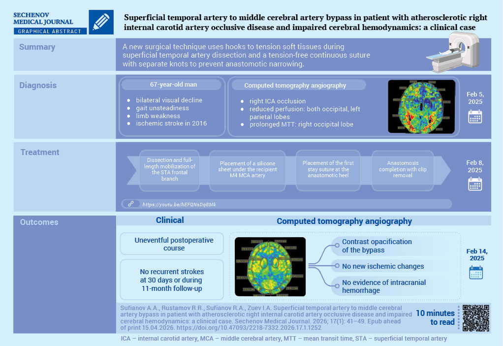
Cerebral revascularization by superficial temporal artery (STA) to middle cerebral artery (MCA) bypass is performed in patients with moyamoya disease, complex aneurysms, and selected extraand intracranial occlusive lesions to augment cerebral perfusion and potentially reduce the risk of ischemic complications and death.
Case report. A 67-year-old patient presented with severe visual impairment (mainly on the right), gait unsteadiness, episodic subjective limb weakness, and marked fatigue. He had a significant medical history, having suffered an ischemic stroke in 2016 in the territory of the right MCA. A computed tomography angiography demonstrated occlusion of the right internal carotid artery and reduced cerebral blood flow in both occipital lobes and the left parietal lobe. An STA-MCA bypass anastomosis was performed. The postoperative course was uneventful; follow-up computed tomography angiography confirmed bypass patency without intracranial hemorrhage or new ischemic lesions, and a 10–15% increase in the cerebral blood volume index (up to 8.6 mL/100 g). No recurrent strokes were observed within 30 days and during 11 months of follow-up.
Discussion. Creation of an STA-MCA anastomosis may offer prospects for improving quality of life after ischemic stroke, including potential amelioration of post-stroke depression and other associated emotional disturbances.
Torkildsen ventriculocisternostomy was historically one of the principal surgical treatments for obstructive hydrocephalus. However, nowadays it tends to be regarded mainly as a salvage procedure when standard shunting or endoscopic ventriculostomy is not feasible.
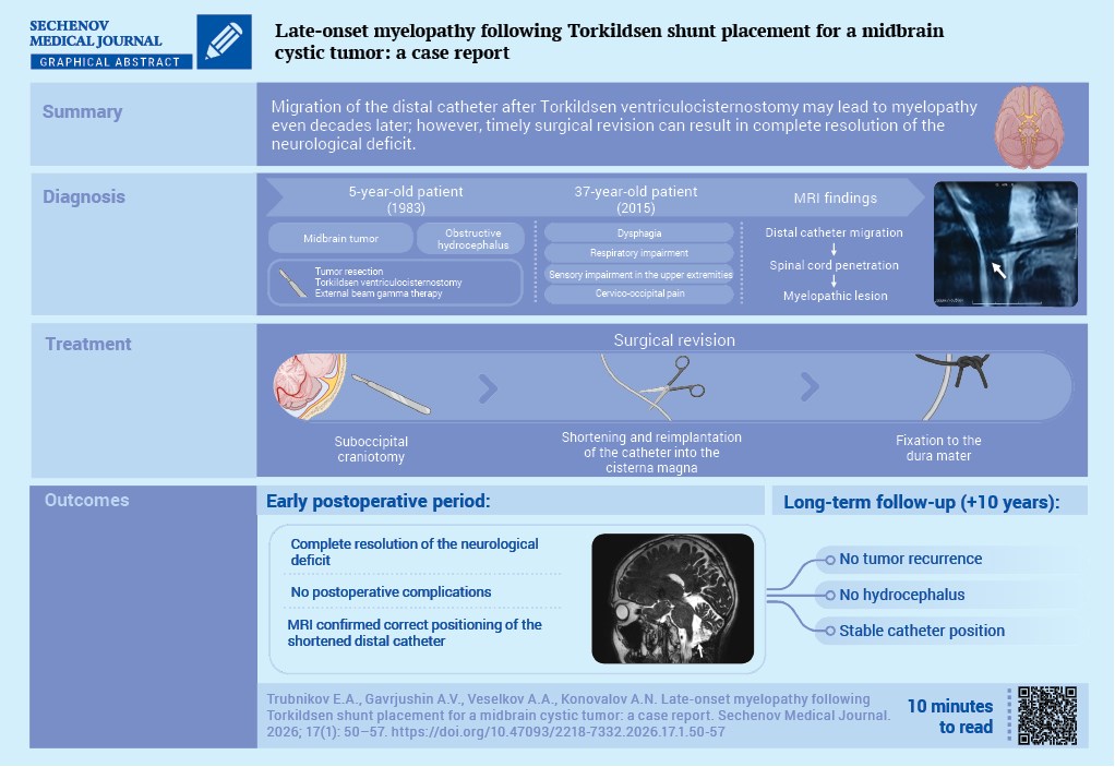
Case report. A 5-year-old boy with obstructive hydrocephalus secondary to a cystic midbrain tumor underwent tumor resection combined with Torkildsen ventriculocisternostomy. Postoperatively, adjuvant radiotherapy was administered, resulting in long-term disease stabilization. At the age of 37 years, 32–33 years after surgery, he developed dysphagia, respiratory disturbances, cervico-occipital pain, and sensory impairment in the upper limbs. Magnetic resonance imaging demonstrated migration of the distal catheter tip with penetration into the upper cervical spinal cord segments and formation of a focal myelopathic lesion. A suboccipital craniotomy was performed; the migrated catheter segment was removed, the system was shortened, and the distal end was reimplanted into the cisterna magna with fixation to the dura mater. Complete regression of neurological deficits was achieved, with a favorable 10-year follow-up.
Discussion. We report a rare delayed case of myelopathy caused by migration of the cisternal catheter tip more than 30 years after Torkildsen ventriculocisternostomy. This observation highlights the need for lifelong surveillance of patients who have undergone such procedures, strict adherence to surgical technique (appropriate catheter length selection and secure fixation), and timely surgical revision at the earliest signs of brainstem dysfunction or involvement of the upper cervical spinal cord.

ISSN 2658-3348 (Online)


