



Contents
Scroll to:
 A. V. Shabunin,
M. M. Tavobilov,
A. A. Karpov,
M. N. Aladin,
A. V. Lantsynova,
K. A. Abramov,
E. B. Kudryash
A. V. Shabunin,
M. M. Tavobilov,
A. A. Karpov,
M. N. Aladin,
A. V. Lantsynova,
K. A. Abramov,
E. B. Kudryash https://doi.org/10.47093/2218-7332.2025.16.4.4-19
Scroll to:
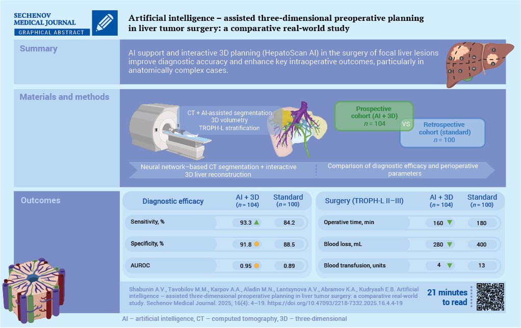
Aim. Evaluation of the diagnostic and clinical effectiveness of the HepatoScan AI system, integrating neural network– based computed tomography (CT) analysis and interactive three-dimensional (3D) preoperative modeling in realworld clinical practice.
Materials and methods. A single-center comparative study was conducted, including a prospective cohort with artificial intelligence-assisted (AI) preoperative planning (n = 104) and a purposefully matched retrospective cohort undergoing standard preoperative planning (n = 100). In the AI-assisted group, automated liver and lesion segmentation, 3D volumetry, and stratification of anatomical complexity using the TROPH-L (Tumor – Regional capsule – Outflow veins – Portal vein – Hepatic bile – Localization) classification were applied. Diagnostic performance was assessed at the patient level using sensitivity, specificity, and AUROC (area under the receiver operating characteristic curve), with histopathological confirmation for malignant tumors and expert interpretation of multiphasic CT for benign lesions. Clinical effectiveness was analyzed in patients classified as TROPH-L II–III.
Results. The compared groups were well balanced in terms of demographic and clinical characteristics, lesion parameters, and distribution of TROPH-L categories. In the overall cohort, the use of HepatoScan AI was associated with a higher sensitivity of preoperative diagnosis compared with the standard approach (93.3% vs. 84.2%; p = 0.008), while maintaining high specificity. The AUROC was higher in the AI group (0.954 vs. 0.892), although the difference was not statistically significant; a similar trend was observed across nosological subgroups. Among patients classified as TROPH-L II–III (n = 54 in the AI group and n = 55 in the standard planning group), AI-assisted planning was associated with a shorter operative time (160 vs. 180 minutes; p = 0.01) and reduced intraoperative blood loss (280 vs. 400 mL; p = 0.004). In addition, a higher rate of R0 resections (100% vs. 91.3%) and lower rates of postoperative complications and in-hospital mortality were observed in the AI group, although these differences were not statistically significant.
Conclusion. Integration of the HepatoScan AI system with interactive 3D preoperative planning is associated with improved diagnostic performance and favorable intraoperative metrics in patients with liver tumors, particularly in anatomically complex cases (TROPH-L II–III). These findings highlight the strong potential of the proposed digital technologies to optimize preoperative planning and warrant further prospective multicenter validation.
Liver tumors represent a major global healthcare challenge and remain one of the leading causes of cancer-related mortality worldwide [1]. In routine clinical practice, modern tertiary care centers are increasingly required to make rapid, accurate, and evidence-based diagnostic and therapeutic decisions when managing patients with focal liver lesions [2]. This group of diseases remains particularly complex: radical treatment is often associated with a high risk of intraoperative and postoperative complications, while long-term outcomes are frequently suboptimal [3].
Importantly, comprehensive diagnostic assessment and surgical planning are required not only for malignant tumors but also for benign focal liver lesions. Large hemangiomas, adenomas, focal nodular hyperplasia (FNH), and complex cystic lesions may, in selected cases, cause significant pain, carry a risk of rupture, or lead to compression of vascular and biliary structures. As a result, these conditions can become clinically relevant and necessitate detailed preoperative evaluation [4].
In this context, there has been a growing interest in the integration of artificial intelligence (AI) technologies and three-dimensional (3D) modeling into clinical hepatobiliary surgery [5]. The combined use of AI-based tools and 3D visualization fundamentally transforms clinical decision-making by shifting it from subjective image interpretation toward objective, quantitative data analysis [6]. Contemporary deep learning algorithms have already demonstrated the ability to automatically segment hepatic structures on contrast-enhanced computed tomography (CT) images with an accuracy of up to 92–94%, improving diagnostic reproducibility while significantly reducing interpretation time [7].
The synergistic application of AI services and 3D modeling enables the creation of highly accurate, patient-specific virtual reconstructions of the liver, allowing detailed visualization of focal lesions and their anatomical relationships with vascular structures and the biliary tree [8]. This approach contributes to a more efficient diagnostic workflow, reducing the need for certain invasive procedures and decreasing the overall burden on the patient [9]. Clinical studies have shown that the use of 3D navigation and reconstruction techniques is associated with a significant reduction in operative time and a lower incidence of R1 resections, thereby directly improving the safety and oncological quality of surgical interventions [4][10]. Consequently, digital technologies not only streamline the diagnostic and planning stages but also enhance the overall quality of surgical care for patients with liver tumors [11].
Objective of the study: to evaluate the diagnostic and clinical effectiveness of the HepatoScan AI clinical decision support system which integrates neural network–based medical image analysis with interactive 3D visualization, in real-world clinical practice.
The development and validation of the clinical decision support system HepatoScan AI for focal liver lesions was carried out in collaboration between Botkin Hospital and the Research and Practical Clinical Center for Diagnostics and Telemedicine Technologies. The system is officially registered in the Russian Federation as computer software [12].
A detailed description of the training process and the performance metrics for the classification of benign and malignant liver lesions has been published previously [13].
To standardize the description of morphological and anatomical characteristics of focal liver lesions and to unify preoperative planning, we developed the TROPH-L classification (Tumor – Regional capsule – Outflow veins – Portal vein – Hepatic bile – Localization). This classification is designed for the systematic analysis of spatial relationships between the tumor and the major hepatic structures, based on data derived from 3D modeling and neural network–based segmentation.
The TROPH-L classification integrates morphological and topographic–anatomical features obtained from patient-specific virtual 3D models with clinically relevant resectability parameters. The classification criteria include:
For preoperative planning, a dedicated 3D modeling workflow was used, including the calculation of volumetric liver parameters and subsequent patient stratification according to the TROPH-L classification. Contrast-enhanced preoperative CT scans were imported into the 3D Slicer software environment in NIfTI format. At this stage, anatomical reconstruction of the liver was performed with semi-automatic segmentation of vascular and biliary structures [14].
In the second stage, the Slicer-Liver extension was applied to automatically delineate Couinaud liver segments and to calculate centerlines of the vascular trees. This was followed by virtual liver resection planning. Using the Resections module, the proposed resection plane was defined, with automatic calculation of hepatic parenchymal volumes, including total liver volume (TLV), resection volume (RV), remnant liver volume (RLV), and tumor volume (TuV).
The relative future liver remnant volume (aFLR, adjusted future liver remnant) was calculated automatically, reflecting the proportion of functionally preserved parenchyma and serving as a key safety criterion for the planned intervention (optimal threshold ≥ 30%). The finalized 3D models were exported in STL format, underwent post-processing in Blender software (Blender Online Community, version 4.0.2) with color labeling of anatomical structures, and were subsequently converted into glTF (.glb) format for use in 3D visualization and augmented reality systems1.
The entire 3D modeling workflow was performed by surgeons from the specialized hepatopancreatobiliary surgery department with experience in 3D modeling, in close collaboration with a radiologist from Botkin Hospital. The initial automatic CT segmentation generated by the HepatoScan AI system was reviewed and, when necessary, manually corrected by the head of the radiology department (Maxim P. Onishchenko).
Final construction of the virtual liver models and determination of the planned RV were conducted through close interdisciplinary collaboration between the operating surgeons (Mikhail M. Tavobilov, Alexey A. Karpov, Aysa V. Lantsynova, Mark N. Aladin, Kirill A. Abramov, Evgeny B. Kudryash) and the 3D modeling specialist (Mark N. Aladin). The average time required to generate an individualized 3D liver model was approximately one hour (Fig. 1).

FIG. 1. Workflow of three-dimensional preoperative modeling in the surgical management of focal liver lesions.
Note: CT – computed tomography; TROPH-L – Tumor – Regional capsule – Outflow veins – Portal vein – Hepatic bile – Localization; 3D – three-dimensional.
The proposed TROPH-L classification enables structured characterization of the morphologic and anatomical features of focal liver lesions and allows stratification of patients into three categories of surgical complexity, which is of critical importance for determining the extent of resection and selecting the optimal treatment strategy (Table 1).
Table 1. Surgical complexity categories according to the TROPH-L classification
Characteristic | I (low risk) | II (moderate risk) | III (high risk) |
Lesion size and growth pattern | <5 cm, peripheral location | 5–10 cm | centrally located or multifocal tumor |
Relationship of the lesion to the hepatic capsule | subcapsular | subcapsular or intraparenchymal | subcapsular or intraparenchymal |
Hepatic vein involvement | none | invasion of segmental veins | invasion of major hepatic vein branches |
Portal vein branch involvement | none | invasion of segmental branches | invasion of major portal vein branches |
Biliary hypertension | none | segmental or sectoral hypertension | lobal hypertension |
Surgical planning | technically safe RV | use of 3D modeling to define the resection plane | detailed 3D reconstruction and assessment of the aFLR |
Volumetric assessment | not required | calculation recommended: TLV, RV, RLV, TuV, aFLR | mandatory calculation: TLV, RV, RLV, TuV, aFLR |
Segmentation example |  |  |  |
Coding example | Tc1R1O0P0H0 – L4b | Ti4R0O1P2H1 – L7 | Ti3R1O3P3H3R – L4–L7 |
Notes: green indicates the biliary ducts, purple represents the hepatic venous system, blue corresponds to the portal venous system, and yellow denotes the focal lesion.
aFLR – adjusted future liver remnant; RLV – remnant liver volume; RV – resection volume; TLV – total liver volume; TuV – tumor volume; 3D – three-dimensional.
This stratification makes it possible to anticipate the scope and technical complexity of surgery, the potential need for extended or combined resections, and the expected risk of postoperative complications. Integration of the TROPH-L classification with the HepatoScan AI system and 3D modeling algorithms provides a comprehensive preoperative planning framework based on objective anatomical and functional data. This approach ensures greater accuracy and consistency of surgical decision-making compared with traditional, largely subjective assessment methods.
A single-center comparative real-world study including a prospective AI-assisted cohort and a purposively matched retrospective cohort treated with conventional preoperative planning.
The required sample size was calculated based on the primary endpoint–differences in diagnostic performance, specifically sensitivity for detection and differential diagnosis of focal liver lesions. According to published data, the implementation of AI algorithms in radiological diagnostics is associated with an 8–12% increase in diagnostic sensitivity. Assuming an expected sensitivity improvement of at least 8%, a two-sided significance level of α = 0.05, and a study power of 80%, the minimum required sample size was estimated to be no fewer than 90 patients per group.
All patients who underwent surgical treatment in the specialized Hepatopancreatobiliary Surgery Department of Botkin Hospital between January 21, 2021, and June 27, 2025, were screened for eligibility.
Inclusion criteria:
Exclusion criteria:
The prospective AI-assisted cohort included patients treated between February 1, 2023, and June 27, 2025, in which the HepatoScan AI clinical decision support system integrated with 3D modeling was used during preoperative planning. Initially, 148 patients were assessed for inclusion; 44 patients met the exclusion criteria. A total of 104 patients were included in the final analysis (Fig. 2). The distribution by pathology was as follows: liver carcinomas (n = 30), including cholangiocarcinoma (CCA, n = 11) and hepatocellular carcinoma (HCC, n = 19), liver hemangiomas (n = 22), simple liver cysts (n = 30), and FNH (n = 22).

FIG. 2. Flow diagram of patient inclusion in the study.
Note: AI – artificial intelligence; CT – computed tomography; TROPH-L – Tumor – Regional capsule – Outflow veins – Portal vein – Hepatic bile – Localization; FNH – focal nodular hyperplasia.
To minimize the impact of differences in pathological composition on diagnostic and surgical outcomes, a purposefully matched retrospective cohort was formed in accordance with established methodology for comparative surgical studies. Matching was performed at the subgroup level according to lesion type, with consideration of lesion size and key demographic characteristics.
For inclusion in the standard preoperative planning group (retrospective cohort), 174 patients with focal liver lesions who underwent surgical treatment between January 21, 2021, and January 25, 2023, were evaluated. Seventy-four patients were excluded based on the exclusion criteria. The final retrospective cohort consisted of 100 patients with the following diagnoses: liver carcinomas (n = 27), including CCA (n = 7) and HCC (n = 20), liver hemangiomas (n = 23), simple liver cysts (n = 30), and FNH (n = 20). Patient inclusion and allocation are illustrated in Figure 2.
All patients from both cohorts were included in the analysis of diagnostic effectiveness. For each patient, a unified preoperative diagnostic conclusion regarding the nature of the focal liver lesion was established and used to calculate diagnostic performance metrics. In patients with multiple lesions, analysis was performed at the level of the clinically dominant lesion that determined the surgical strategy.
Comparison of HepatoScan AI–assisted results with the reference standard was performed by expert assessment at the level of the final clinical diagnostic conclusion.
The reference standard varied depending on lesion type. For suspected malignant tumors, diagnostic accuracy was assessed by comparing the preoperative HepatoScan AI conclusion with results of preoperative morphological verification (biopsy), followed by confirmation of the final diagnosis based on histopathological examination of the resected specimen. For benign focal liver lesions, the reference standard was the conclusion of an experienced radiologist based on multiphasic contrast-enhanced CT, corroborated by intraoperative findings and the clinical course of the disease.
The analysis of clinical effectiveness of the HepatoScan AI clinical decision support system included patients from both cohorts with anatomically complex focal liver lesions corresponding to TROPH-L categories II–III.
Patients with TROPH-L category I lesions were excluded from this analysis: 50 patients from the prospective cohort and 45 patients from the retrospective cohort. All patients with simple liver cysts in both cohorts were excluded; all of them underwent cyst fenestration.
The final clinical effectiveness analysis included 54 patients from the prospective cohort and 55 patients from the retrospective cohort with liver carcinomas, hemangiomas, and FNH. The compared groups were evaluated with respect to operative time, intraoperative blood loss, and the rate of blood transfusions. In addition, oncological radicality in patients with liver carcinomas (rate of R0 resections), postoperative complication rates – including severe complications graded III–V according to the Clavien–Dindo classification – length of hospital stay, and in-hospital mortality were analyzed.
All surgical procedures were performed using standard surgical techniques. The choice of surgical approach (open or laparoscopic) was determined on an individual basis, taking into account lesion localization, size, relationships with vascular and biliary structures, and the planned extent of resection. Liver parenchymal transection was carried out using an ultrasonic dissector and bipolar coagulation; in selected cases, an ultrasonic aspirator–destructor was employed with stepwise identification and control of vascular and biliary structures. The Pringle maneuver was applied selectively and only when indicated, in cases with an anticipated risk of significant intraoperative blood loss.
Statistical analysis was performed using IBM SPSS Statistics software version 30.0 (SPSS: An IBM Company, USA) and the R programming language (version 4.4.1), with the use of specialized packages pROC (version 1.18.0) and stats (base R package).
Quantitative variables were initially assessed for normality using the Shapiro–Wilk test. Depending on the distribution, data are presented as mean with standard deviation (M ± SD) or median with interquartile range (Me (Q1; Q3)). Between-group comparisons of continuous variables were performed using Student’s t-test for normally distributed data or the Mann–Whitney U test for non-normally distributed data.
Categorical variables are presented as absolute and relative frequencies and were compared using Pearson’s χ² test or Fisher’s exact test when expected cell counts were below five. Comparisons of blood transfusion rates between groups were performed using aggregated data and a Poisson regression model to calculate incidence rate ratios (IRR).
Diagnostic performance of the HepatoScan AI system was evaluated on a per-patient basis and included calculation of sensitivity, specificity, and the area under the ROC curve (AUROC), with corresponding 95% confidence intervals (CI).
For malignant focal liver lesions, the reference standard was based on preoperative morphological verification (biopsy) followed by confirmation of the final diagnosis through histopathological examination of the resected specimen. For benign focal liver lesions, the reference standard consisted of the radiologist’s conclusion based on multiphasic contrast-enhanced CT, corroborated by intraoperative findings and the clinical course of the disease. Comparisons of AUROC values between groups were performed using the nonparametric DeLong test.
Differences were considered statistically significant at p < 0.05.
The study groups did not differ significantly with respect to key demographic characteristics, including age, sex, and the presence of comorbid or background diseases. No statistically significant differences were observed between groups in terms of lesion size, number, multiplicity, or anatomical localization. Retrospective assessment of case complexity in the standard preoperative planning group using the TROPH-L classification demonstrated a distribution of risk categories comparable to that of the prospective AI-assisted preoperative planning group (Table 2).
Table 2. Baseline characteristics of patients in the study groups
Parameter | AI-assisted group (n = 104) | Conventional planning group (n = 100) | p value |
Age, years | 59.7 ± 11.1 | 60.3 ± 10.5 | 0.62 |
Male / female | 56 / 44 (54% / 46%) | 53 / 47 (53% / 47%) | 0.67 |
BMI, kg/m² | 27.1 (24.4; 29.8) | 26.8 (24.0; 29.6) | 0.51 |
Arterial hypertension | 52 (50%) | 49 (49%) | 0.89 |
Type 2 diabetes mellitus | 21 (20.2%) | 19 (19%) | 0.83 |
Chronic liver disease | 10 (9.6%) | 13 (13%) | 0.45 |
Cardiovascular diseases | 30 (28.8%) | 28 (28%) | 0.89 |
ASA physical status, class | |||
I–II | 64 (61.5%) | 58 (58%) | 0.61 |
III | 40 (38.5%) | 42 (42%) | |
Lesion diameter, mm | 65 (45; 85) | 60 (43; 77) | 0.61 |
Multiple lesions (≥ 2) | 26 (25%) | 20 (20%) | 0.39 |
Lesion localization | |||
right lobe | 62 (60%) | 65 (65%) | 0.71 |
left lobe | 31 (30%) | 25 (25%) | |
both lobes | 11 (10%) | 10 (10%) | |
TROPH-L categories | |||
I | 50 (48%) | 45 (45%) | 0.90 |
II | 40 (38.5%) | 40 (40%) | |
III | 14 (13.5%) | 15 (15%) | |
Carcinomas, stage | n = 30 | n = 27 | |
I | 12 (40%) | 10 (37%) | 0.97 |
II | 13 (43%) | 12 (44%) | |
III | 5 (17%) | 5 (19%) | |
Carcinomas, histological grade | n = 30 | n = 27 | |
high | 9 (30%) | 8 (30%) | 0.98 |
medium | 15 (50%) | 13 (48%) | |
low | 6 (20%) | 6 (22%) |
Notes: quantitative variables are presented as mean with standard deviation (M ± SD) or median with interquartile range (Me (Q1; Q3)), categorical variables are presented as the absolute number of patients with the characteristic and the proportion within the group, expressed as a percentage (in parentheses).
AI – artificial intelligence; ASA – American Society of Anesthesiologists; BMI – body mass index; TROPH-L – Tumor – Regional capsule – Outflow veins – Portal vein – Hepatic bile – Localization.
When individual nosological subgroups were analyzed, no statistically significant differences in sensitivity, specificity, or AUROC of the ROC curve were observed between the groups. Across all subgroups, a consistent trend toward higher diagnostic performance metrics was noted in the AI-assisted preoperative planning group; however, due to the limited size of the subgroups, these differences did not reach statistical significance. When diagnostic metrics were compared across all types of focal liver lesions, integration of the HepatoScan AI system was associated with improved diagnostic sensitivity for focal liver lesions compared with the standard diagnostic approach (Table 3).
Table 3. Diagnostic performance of the HepatoScan AI system in determining the nature of focal liver lesions
AI-assisted group (n = 104) | Conventional planning group (n = 100) | p value | |
Carcinoma | n = 30 | n = 27 | |
sensitivity | 92.9 (78.7–98.2) | 81.5 (63.3–91.8) | n.s. |
specificity | 93 (85.1–97.1) | 90 (81.5–95.3) | n.s. |
AUROC | 0.95 (0.89–1.00) | 0.88 (0.79–0.97) | n.s. |
Focal nodular hyperplasia | n = 22 | n = 20 | |
sensitivity | 88.2 (66.7–95.3) | 78.6 (58.4–91.9) | n.s. |
specificity | 90.5 (81.9–95.0) | 88 (78.5–93.1) | n.s. |
AUROC | 0.93 (0.85–1.00) | 0.87 (0.77–0.97) | n.s. |
Hemangioma | n = 22 | n = 23 | |
sensitivity | 96.3 (78.2–99.2) | 85 (67.9–95.5) | n.s. |
specificity | 94.2 | 89.5 (80.8–94.6) | n.s. |
AUROC | 0.97 (0.92–1.00) | 0.90 (0.81–0.99) | n.s. |
Simple liver cyst | n = 30 | n = 30 | |
sensitivity | 98.1 (83.3–99.4) | 92 (78.7–98.2) | n.s. |
specificity | 95.3 (86.9–97.9) | 90.8 (82.5–96.0) | n.s. |
AUROC | 0.98 (0.94–1.00) | 0.93 (0.86–1.00) | n.s. |
All types of focal liver lesions | |||
sensitivity | 93.3 (88.2–97.1) | 84.2 (77.1–90.3) | 0.008 |
specificity | 91.8 (86.7–96.4) | 88.5 (82.4–93.7) | n.s. |
AUROC | 0.95 (0.90–1.00) | 0.89 (0.81–0.98) | n.s. |
Notes: sensitivity and specificity data are presented as percentages with 95% CI (in parentheses), and AUROC as a value with a 95% CI (in parentheses).
AI – artificial intelligence; AUROC – area under the receiver operating characteristic curve; CI – confidence interval; n.s. – not significant.
The use of 3D modeling was most clinically justified in patients classified as TROPH-L categories II and III. This subgroup included 54 patients from the prospective cohort and 55 patients from the retrospective cohort. The compared groups did not differ significantly in terms of key clinical and demographic characteristics, prevalence of comorbidities, lesion size, anatomical localization, lesion multiplicity, or – among patients with carcinomas – tumor stage and histological grade (Table 4).
Table 4. Baseline characteristics of patients in the study groups with TROPH-L complexity categories II–III
Parameter | AI-assisted group (n = 54) | Conventional planning group (n = 55) | p value |
Age, years | 60.5 ± 10.2 | 61.2 ± 9.8 | n.s. |
Male / female | 29 / 25 (54% / 46%) | 29 / 26 (53% / 47%) | n.s. |
BMI, kg/m² | 27.4 (24.5; 31.2) | 27.0 (24.0; 30.8) | n.s. |
Arterial hypertension | 27 (50%) | 27 (49%) | n.s. |
Type 2 diabetes mellitus | 11 (20%) | 10 (18%) | n.s. |
Chronic liver disease | 5 (9%) | 7 (13%) | n.s. |
Cardiovascular diseases | 16 (30%) | 15 (27%) | n.s. |
ASA physical status, class | |||
I–II | 33 (61%) | 32 (58%) | n.s. |
III | 21 (39%) | 23 (42%) | |
Lesion diameter, mm | 75 (55; 95) | 70 (50; 90) | n.s. |
Multiple lesions (≥ 2) | 17 (32%) | 14 (25.5%) | n.s. |
Lesion localization | |||
right lobe | 32 (59%) | 36 (65.5%) | |
left lobe | 16 (30%) | 14 (25.5%) | n.s. |
both lobes | 6 (11%) | 5 (9%) | |
TROPH-L categories | |||
II | 40 (74%) | 40 (73%) | n.s. |
III | 14 (26%) | 15 (27%) | |
Carcinomas, stage | n = 26 | n = 23 | |
I | 8 (31%) | 6 (26%) | |
II | 13 (50%) | 12 (52%) | n.s. |
III | 5 (19%) | 5 (22%) | |
Carcinomas, histological grade | n = 26 | n = 23 | |
high | 8 (31%) | 7 (30%) | |
medium | 13 (50%) | 11 (48%) | n.s. |
low | 5 (19%) | 5 (22%) |
Notes: quantitative variables are presented as mean with standard deviation (M ± SD) or median with interquartile range (Me (Q1; Q3)), categorical variables are presented as the absolute number of patients with the characteristic and the proportion within the group, expressed as a percentage (in parentheses).
AI – artificial intelligence; ASA – American Society of Anesthesiologists; BMI – body mass index; n.s. – not significant; TROPH-L – Tumor – Regional capsule – Outflow veins – Portal vein – Hepatic bile – Localization.
At the surgical stage, integration of the neural network–based clinical decision support system HepatoScan AI in combination with 3D preoperative modeling had a meaningful impact on the choice of surgical strategy in patients classified as TROPH-L categories II–III. In the AI-assisted preoperative planning group, a trend toward a more parenchyma-sparing approach was observed: extended anatomical liver resections were performed less frequently (27.8% vs 38.2% in the standard preoperative planning group), while the proportion of non-anatomical (atypical) resections was higher (55.6% vs 45.5%). The frequency of segmentectomies did not differ significantly between groups (Table 5).
Table 5. Clinical effectiveness of the HepatoScan AI system and three-dimensional preoperative modeling
Characteristic | AI-assisted group (n = 54) | Conventional planning group (n = 55) | p value |
Type of surgical procedure | |||
extended anatomical liver resection | 15 (28%) | 21 (38%) | |
non-anatomical (atypical) resection | 30 (56%) | 25 (46%) | n.s. |
segmentectomy | 9 (17%) | 9 (16%) | |
Operative time, min | 160 (135; 190) | 180 (150; 210) | 0.01 |
Intraoperative blood loss, mL | 280 (200; 400) | 400 (300; 600) | 0.004 |
Blood transfusions, number of packed red blood cell units, n | 4 | 13 | 0.04 |
R0 resection for liver carcinomas, number of patients | 26 out of 26 (100%) | 21 out of 23 (91%) | n.s. |
All postoperative complications, number of patients | 15 (28%) | 22 (40%) | n.s. |
Severe complications (Clavien–Dindo grades III–V), number of patients | 5 (9%) | 10 (18%) | n.s. |
Length of hospital stay, days | 11 (9; 13) | 12 (10; 15) | n.s. |
In-hospital mortality, number of patients | 0 | 2 (3.6%) | n.s. |
Notes: quantitative variables are presented as median with interquartile range (Me (Q1; Q3)), categorical variables are presented as the absolute number of patients with the characteristic and the proportion within the group, expressed as a percentage (in parentheses).
AI – artificial intelligence; n.s. – not significant.
In the subgroup of patients with carcinomas, R0 resection was achieved in all patients in the AI-assisted preoperative planning group (100%) compared with 91.3% in the standard preoperative planning group; however, this difference did not reach statistical significance (p = 0.18).
The use of AI technologies and 3D modeling was associated with improved intraoperative parameters. In the AI-assisted preoperative planning group, the mean operative time was reduced by approximately 20 minutes. A more pronounced effect was observed for intraoperative blood loss: mean blood loss in the AI-assisted group was 120 mL lower than in the standard preoperative planning group. In the AI-assisted preoperative planning group, a total of 4 units of packed red blood cells were transfused in 54 patients, corresponding to 0.07 units per patient, whereas in the standard preoperative planning group 13 units were transfused in 55 patients (0.24 units per patient). Accordingly, the rate of blood transfusion use was approximately 3.2 times higher in the standard preoperative planning group (incidence rate ratio 3.19; 95% CI 1.04–9.76).
The reduction in intraoperative surgical trauma was reflected in postoperative outcomes. In the AI-assisted preoperative planning group, a trend toward a lower overall rate of postoperative complications (28% vs 40%) as well as severe complications (Clavien–Dindo grades III–V: 9% vs 18%) was observed compared with the standard preoperative planning group; however, given the sample size, these differences did not reach statistical significance (p = 0.17).
In the AI-assisted preoperative planning group, severe complications were recorded in five patients and included postoperative bleeding (n = 3), pulmonary embolism (n = 1), and a biliary fistula Grade B according to the International Study Group of Liver Surgery (ISGLS) classification (n = 1) requiring percutaneous drainage under ultrasound guidance. No cases of acute postoperative liver failure were observed in this group. In the standard preoperative planning group, severe postoperative complications (n = 10) included acute postoperative liver failure (n = 3), clinically significant postoperative bleeding (n = 4), pulmonary embolism (n = 1), and biliary complications in the form of ISGLS Grade B biliary fistulas (n = 2), which required percutaneous drainage under ultrasound guidance.
In-hospital mortality was observed only in the standard preoperative planning group (n = 2); in both cases, death was attributable to progression of acute postoperative liver failure following extensive liver resections.
The results of the present study demonstrate that integration of AI technologies into the diagnostic workflow for liver tumors is associated with improved detection of focal liver lesions. Compared with the standard diagnostic approach, use of the HepatoScan AI system resulted in a statistically significant increase in the sensitivity of preoperative diagnosis for all focal liver lesions by 9.1% (from 84.2% to 93.3%), while maintaining a high level of specificity (91.8%).
The observed improvement in diagnostic accuracy with AI assistance is consistent with current literature. Several studies have shown that deep learning models are capable of detecting hepatic tumor lesions with accuracy comparable to expert radiologist assessment and, in some cases, identifying additional lesions not recognized during conventional visual analysis [6][9]. Moreover, it has been reported that the combined use of expert interpretation and AI algorithms increases the sensitivity of focal liver lesion detection from approximately 80% to 88% [15].
Visualization of the liver as an interactive 3D model enables “virtual navigation” within the organ, precise calculation of RV, and prediction of the risk of postoperative liver failure – features that are particularly important in cases of complex tumor localization. The TROPH-L classification developed and first introduced in this study integrates AI-based segmentation with 3D reconstruction and illustrates the feasibility of using objective morphological and topographic–anatomical parameters for standardized stratification of surgical risk. Overall, this approach aligns with the global trend toward personalized surgical treatment based on large-scale data analysis and the application of AI [16].
In the present study, automated 3D reconstruction of liver anatomy demonstrated clear clinical benefits in patients with moderate and high surgical risk, corresponding to TROPH-L categories II–III. Compared with standard preoperative planning, the AI-assisted group showed more favorable intraoperative and postoperative outcomes: operative time was reduced by an average of 11%, intraoperative blood loss was approximately 30% lower, and the need for blood transfusions was reduced by about 3.2-fold. These findings reflect improved selection of surgical approach and extent of resection based on detailed preoperative 3D anatomical analysis.
Our results are consistent with data from multicenter studies demonstrating that the use of 3D visualization in liver resection planning leads to statistically significant reductions in operative time and blood loss, lower rates of postoperative liver failure and other complications, and shorter hospital stays [10][11]. In particular, a recent prospective study showed that preoperative assessment using 3D reconstruction is associated with reduced surgical trauma and faster postoperative recovery compared with standard two-dimensional navigation [11].
The impact of 3D planning on oncological outcomes should also be emphasized. According to published data, the use of 3D navigation facilitates more accurate definition of resection margins and increases the rate of R0 resections [17]. In the present study, no cases of positive surgical margins (R1 resections) were observed in the group undergoing 3D reconstruction, whereas such cases occurred, albeit infrequently, in the standard preoperative planning group (2 of 23 patients, 8.7%). These findings are in agreement with meta-analyses demonstrating that 3D-assisted liver resections are associated with wider resection margins and lower recurrence rates [11][16][17].
Furthermore, more accurate calculation of the aFLR and detailed 3D visualization of tumor relationships with vascular and biliary structures may have contributed to a higher rate of parenchyma-sparing yet oncologically radical resections, avoiding unjustifiably extensive liver resections. This approach was associated with a trend toward a lower incidence of severe postoperative complications (Clavien–Dindo grade ≥ III) and the absence of perioperative mortality, reflecting improved surgical safety [17][18].
Taken together, these findings suggest that integration of neural network–based algorithms and 3D modeling can serve as an effective decision-support tool for personalized surgical planning. Such an approach has the potential to enhance the quality of preoperative diagnosis and improve short-term surgical outcomes in patients with focal liver lesions, making operative planning more objective and reproducible and surgical interventions safer and more effective.
At the same time, widespread implementation of these systems requires further investigation and resolution of several organizational and technical challenges. It should be acknowledged that most studies on AI-based liver imaging are based on retrospective data from relatively small cohorts [19], which limits the strength of conclusions. Nevertheless, the rapid advancement of machine learning technologies and the accumulation of prospective clinical data provide grounds to anticipate an increasingly prominent role for such tools in routine clinical practice.
When interpreting the results of this study, several limitations should be considered. First, the comparison between a retrospective cohort (2021–2023) and a prospective cohort (2023–2025) may reflect not only the impact of digital technology implementation but also temporal changes in surgical techniques and perioperative patient management. Second, the relatively small sample size – particularly after stratification by nosological subgroups and TROPH-L complexity categories – limits the statistical power of the analysis and the ability to detect differences in certain clinical outcomes. In addition, different reference standards were applied for different types of focal liver lesions, and comparisons involving the HepatoScan AI system were performed at the level of the final clinical diagnostic conclusion, which does not fully exclude the influence of expert interpretation. Finally, the single-center design of the study and the use of the proprietary TROPH-L classification may limit the generalizability of the findings and their direct comparability with results from other studies.
In this single-center comparative study, integration of neural network–based CT analysis with interactive 3D preoperative modeling (HepatoScan AI) was associated with increased sensitivity for detection and classification of focal liver lesions compared with the standard approach. Automated segmentation, quantitative 3D volumetry, and the TROPH-L classification enabled standardized assessment of anatomical complexity and resectability, which was particularly relevant for patients classified as TROPH-L categories II–III. In this subgroup, AI assistance and 3D planning were associated with improvements in selected intraoperative parameters and a trend toward a lower incidence of severe postoperative complications, while maintaining oncological radicality. Further prospective multicenter studies are warranted to standardize methodologies for evaluating digital tools in liver surgery and to validate these findings across broader clinical settings.
Alexey V. Shabunin and Mikhail M. Tavobilov conceptualized and designed the study and contributed to the surgical procedures. Mark N. Aladin curated the clinical data, contributed to the surgical procedures, performed 3D image segmentation and post-processing, conducted data analysis, and contributed to the development and validation of artificial intelligence algorithms and 3D modeling. Alexey A. Karpov contributed to the surgical procedures and provided organizational and methodological oversight of the surgical component of the study. Aysa V. Lantsynova and Kirill A. Abramov contributed to the surgical procedures, conducted the literature review, and supported clinical data analysis. Evgeny B. Kudryash contributed to the surgical procedures and performed statistical analysis and interpretation of the results. Alexey V. Shabunin, Mikhail M. Tavobilov, and Mark N. Aladin contributed to manuscript drafting and critical revision. All authors reviewed and approved the final manuscript.
Ethics statements. The study was approved by the Local Ethics Committee (Protocol No. 15 dated 11 October 2022) of the Russian Medical Academy of Continuous Professional Education, Ministry of Health of the Russian Federation.
Data availability. The data confirming the findings of this study are available from the authors upon reasonable request. Data and statistical methods used in the article were examined by a professional biostatistician on the Sechenov Medical Journal editorial staff.
Conflict of interest. The authors declare that there is no conflict of interests.
Financing. This study was supported by a government-funded Research and Development project aimed at reducing postoperative complications and mortality and improving survival outcomes in patients with benign and malignant liver diseases through the use of neural network technologies and preoperative three-dimensional modeling (EGISU No. 123042600026-2). The project was approved by the Moscow City Health Department under Order No. 1196 dated December 21, 2022, covering the period 2023–2025.
Acknowledgements. The authors express their deep gratitude to the Head of the Department of Radiology of Botkin Hospital Maxim P. Onishchenko for his assistance in organizing radiological examinations, as well as to the staff of the Research and Practical Clinical Center for Diagnostics and Telemedicine Technologies: Chief Physician Yuri A. Vasiliev, Head of the Directorate for Scientific Division Management Olga V. Omelyanskaya, Head of the Department of Medical Informatics, Radiomics, and Radiogenomics Kirill M. Arzamasov, Deputy Head of the Department of Medical Informatics, Radiomics and Radiogenomics Lev D. Pestrenin, and Junior Researcher of the Department of Radiomics and Radiogenomics Ekaterina F. Savkina, for their contribution to the development of the HepatoScan AI system and to the processing and analysis of radiological examinations.
1. Blender Foundation. Blender — free and open-source 3D creation suite. Amsterdam: Blender Foundation; 2025. https://www.blender.org (access date: 24.10.2025).
1. Rumgay H., Arnold M., Ferlay J., et al. Global burden of primary liver cancer in 2020 and predictions to 2040. J Hepatol. 2022 Dec; 77(6): 1598–1606. https://doi.org/10.1016/j.jhep.2022.08.021. Epub 2022 Oct 5. PMID: 36208844
2. Ding Z.B., Shi Y.H., Chen J.F., et al. Recent advances in surgical management strategies for hepatocellular carcinoma. hLife. 2024; 2(9): 439–453. https://doi.org/10.1016/j.hlife.2024.06.007
3. Yang S., Ni H., Zhang A., et al. Impact of postoperative morbidity on the prognosis of patients with hepatocellular carcinoma after laparoscopic liver resection: a multicenter observational study. Sci Rep. 2025 Jan; 15(1): 1724. https://doi.org/10.1038/s41598-024-85020-9. PMID: 39799160
4. Rahman H., Bukht T.F.N., Imran A., et al. A Deep Learning Approach for Liver and Tumor Segmentation in CT Images Using ResUNet. Bioengineering (Basel). 2022 Aug; 9(8): 368. https://doi.org/10.3390/bioengineering9080368. PMID: 36004893
5. Chatzikomnitsa P., Gkaitatzi A.D., Papakonstantinou M., et al. The role of artificial intelligence and 3D printing in minimally invasive liver surgery. Mini-invasive Surg. 2025; 9: 8. https://doi.org/10.20517/2574-1225.2024.78
6. Efstathiou A., Charitaki E., Triantopoulou C., Delis S. Artificial Intelligence and Digital Tools Across the Hepato-PancreatoBiliary Surgical Pathway: A Systematic Review. J Clin Med. 2025 Sep; 14(18): 6501. https://doi.org/10.3390/jcm14186501. PMID: 41010705
7. Manjunath R.V., Kwadiki K. Modified U-NET on CT images for automatic segmentation of liver and its tumor. Biomedical Engineering Advances. 2022; 4: 100043. https://doi.org/10.1016/j.bea.2022.100043
8. Banchini F., Capelli P., Hasnaoui A., et al. 3-D reconstruction in liver surgery: a systematic review. HPB (Oxford). 2024 Oct; 26(10): 1205–1215. https://doi.org/10.1016/j.hpb.2024.06.006. Epub 2024 Jun 18. PMID: 38960762
9. Yin C., Zhang H., Du J., et al. Artificial intelligence in imaging for liver disease diagnosis. Front Med (Lausanne). 2025 Apr; 12: 1591523. https://doi.org/10.3389/fmed.2025.1591523. PMID: 40351457
10. Zeng X., Tao H., Dong Y., et al. Impact of three-dimensional reconstruction visualization technology on short-term and long-term outcomes after hepatectomy in patients with hepatocellular carcinoma: a propensity-score-matched and inverse probability of treatment-weighted multicenter study. Int J Surg. 2024 Mar; 110(3): 1663–1676. https://doi.org/10.1097/JS9.0000000000001047. PMID: 38241321
11. Agrawal H., Tanwar H., Gupta N. Revolutionizing hepatobiliary surgery: Impact of three-dimensional imaging and virtual surgical planning on precision, complications, and patient outcomes. Artif Intell Gastroenterol. 2025; 6(1): 106746. https://doi.org/10.35712/aig.v6.i1.106746
12. Васильев Ю.А., Владзимирский А.В., Омелянская О.В. и др. MOSMEDSOFT: СТ HepatoScan AI. Свидетельство о государственной регистрации программы для ЭВМ RU 2025683642. Российская Федерация; заявка № 2025682549; дата регистрации 28.08.2025; дата публикации 05.09.2025. / Vasilyev Yu.A., Vladzimirskiy A.V., Omelyanskaya O.V., et al. MOSMEDSOFT: ST HepatoScan AI. Certificate of state registration of computer software No. RU 2025683642. Russian Federation; 2025 (In Russian).
13. Shabunin А.V., Vasilyev Y.А., Tavobilov М.М., et al. Development of a clinical decision support system for the diagnosis of space-occupying liver lesions using artificial intelligence methods. Annaly khirurgicheskoy gepatologii = Annals of HPB surgery. 2025; 30(2): 23–32 (In Russian). https://doi.org/10.16931/1995-5464.2025-2-23-32. EDN: NKWRDR
14. Fedorov A., Beichel R., Kalpathy-Cramer J., et al. 3D Slicer as an image computing platform for the Quantitative Imaging Network. Magn Reson Imaging. 2012 Nov; 30(9): 1323– 1341. https://doi.org/10.1016/j.mri.2012.05.001. PMID: 22770690
15. Dai H., Xiao Y., Fu C., et al. Deep Learning-Based Approach for Identifying and Measuring Focal Liver Lesions on ContrastEnhanced MRI. J Magn Reson Imaging. 2025 Jan; 61(1): 111– 120. https://doi.org/10.1002/jmri.29404. Epub 2024 Jun 3. PMID: 38826142
16. Mei G., Yu J. Research on CT image segmentation and classification of liver tumors based on attention mechanism and improved U-Net model. Technol Health Care. 2025 Sep; 33(5): 2468–2483. https://doi.org/10.1177/09287329251329294. Epub 2025 Apr 30. PMID: 40302502
17. Montalti R., Rompianesi G., Cassese G., et al. Role of preoperative 3D rendering for minimally invasive parenchyma sparing liver resections. HPB. 2023; 25(8): 915–923. https://doi.org/10.1016/j.hpb.2023.04.008
18. Hori M., Suzuki Y., Sofue K., et al. Artificial intelligence in imaging diagnosis of liver tumors: current status and future prospects. Abdom Radiol (NY). 2025 Jun. https://doi.org/10.1007/s00261-025-05059-8. Epub ahead of print. PMID: 40536541
19. Li L., Cheng S., Li J., et al. Randomized comparison of AI enhanced 3D printing and traditional simulations in hepatobiliary surgery. NPJ Digit Med. 2025 Jun; 8(1): 293. https://doi.org/10.1038/s41746-025-01571-9. PMID: 40457016
Alexey V. Shabunin, Dr. of Sci. (Medicine), Professor, Academician of the RAS, Head of Department of Surgery, Transplantology and Applied Oncology; Director
5, 2nd Botkinsky proezd, Moscow, 125284; 2/1, bld. 1, Barrikadnaya str., Moscow, 125993
Mikhail M. Tavobilov, Dr. of Sci. (Medicine), Professor, Department of Surgery, Transplantology and Applied Oncology; Head of the Department of Hepatopancreatobiliary Surgery
5, 2nd Botkinsky proezd, Moscow, 125284; 2/1, bld. 1, Barrikadnaya str., Moscow, 125993
Alexey A. Karpov, Dr. of Sci. (Medicine), Associate Professor, Department of Surgery, Transplantology and Applied Oncology; Senior Researcher, Deputy Chief Physician for Surgery
5, 2nd Botkinsky proezd, Moscow, 125284; 2/1, bld. 1, Barrikadnaya str., Moscow, 125993
Mark N. Aladin, Junior Researcher, Surgeon, Department of Hepatopancreatobiliary Surgery
5, 2nd Botkinsky proezd, Moscow, 125284
Aysa V. Lantsynova, Cand. of Sci. (Medicine), Junior Researcher, Surgeon, Department of Hepatopancreatobiliary Surgery
5, 2nd Botkinsky proezd, Moscow, 125284
Kirill A. Abramov, Cand. of Sci. (Medicine), Junior Researcher, Surgeon, Department of Hepatopancreatobiliary Surgery
5, 2nd Botkinsky proezd, Moscow, 125284
Evgeny B. Kudryash, postgraduate student, Department of Surgery, Transplantology and Applied Oncology; Junior Researcher, Surgeon, Department of Hepatopancreatobiliary Surgery
5, 2nd Botkinsky proezd, Moscow, 125284; 2/1, bld. 1, Barrikadnaya str., Moscow, 125993

|
1. Checklist MedinAI for reporting artificial intelligence studies in medicine | |
| Subject | ||
| Type | Research Instrument | |
Download
(4MB)
|
Indexing metadata ▾ | |
Журнал «Сеченовский вестник» |
| Sechenov Medical Journal |
Рецензии на рукопись |
| Peer-review reports |
Название / Title | Искусственный интеллект в сочетании с трехмерным предоперационным планированием в хирургии опухолей печени: сравнительное исследование в реальной клинической практике / Artificial intelligence – assisted three-dimensional preoperative planning in liver tumor surgery: a comparative real-world study
|
Раздел / Section
| ХИРУРГИЯ/ SURGERY
|
Тип / Article | Оригинальная статья / Original article |
Номер / Number | 1369
|
Страна/территория / Country/Territory of origin | Россия / Russia |
Язык / Language | Русский / Russian Английский / English
|
Источник / Manuscript source | Инициативная рукопись / Unsolicited manuscript |
Дата поступления / Received | 16.10.2025 |
Тип рецензирования / Type ofpeer-review | Двойное слепое / Double blind |
Язык рецензирования / Peer-review language | Русский / Russian
|
РЕЦЕНЗЕНТ А / REVIEWER A
Инициалы / Initials | 1369_А
|
Научная степень / Scientific degree | Доктор медицинских наук / Dr. of Sci. (Medicine)
|
Страна/территория / Country/Territory | Россия / Russia
|
Дата рецензирования / Date of peer-review | 29.11.2025 |
Число раундов рецензирования / Number of peer-review rounds | 2 |
Финальное решение / Final decision | принять к публикации / accept
|
ПЕРВЫЙ РАУНД РЕЦЕНЗИРОВАНИЯ / FIRST ROUND OF PEER-REVIEW
Представленная к рецензированию статья посвящена актуальной и практически значимой проблеме – внедрению цифровых технологий, а именно искусственного интеллекта (ИИ) и трёхмерного (3D) моделирования, в комплексный процесс диагностики и хирургического лечения пациентов с очаговыми образованиями печени. Работа носит комплексный характер, включая разработку программного обеспечения (HepatoScan AI), создание алгоритма 3D-планирования, предложение оригинальной классификации TROPH-L и их клиническую апробацию.
Актуальность исследования не вызывает сомнений. Новизна работы значительна. Авторы не просто применяют существующие инструменты, а представляют собственную систему поддержки принятия решений (HepatoScan AI) и оригинальную классификацию TROPH-L для стратификации риска.
Исследование проведено на выборке в 204 пациента, которая формально является репрезентативной, однако возникает вопрос по поводу показаний для хирургического лечения кист и гемангиом печени, составляющих подавляющую часть нозологий; необходимо отдельно и подробно обосновать показания для хирургического лечения этих пациентов, так как в настоящее время хирургическое лечение этих заболеваний выполняется достаточно редко.
Дизайн исследования (сравнение ретроспективной и проспективной когорт) в целом адекватен для оценки эффективности нового метода. Использованная литература актуальна.
В процессе рецензирования возникли следующие замечания/комментарии:
В целом, статья является серьезной, научно обоснованной и практико-ориентированной работой. Авторы продемонстрировали не только возможность, но и определенные преимущества интеграции цифровых технологий в реальную клиническую практику хирургии печени. Полученные результаты имеют высокую ценность для отечественного здравоохранения. После устранения указанных недостатков необходимо повторное рецензирование статьи.
ЗАКЛЮЧЕНИЕ: переработка и повторное рецензирование.
ВТОРОЙ РАУНД РЕЦЕНЗИРОВАНИЯ/ SECOND ROUND OF PEER-REVIEW
Авторы подготовили обновленный вариант статьи с внесенными исправлениями в соответствии с замечаниями. В представленном варианте в статье устранены недочеты, даны пояснения по неоднозначным аспектам (см. первичную рецензию).
Рукопись может быть принята к публикации.
РЕЦЕНЗЕНТ B / REVIEWER B
Инициалы / Initials | 1369_В
|
Научная степень / Scientific degree | Доктор медицинских наук / Dr. of Sci. (Medicine)
|
Страна/территория / Country/Territory | Россия / Russian
|
Дата рецензирования / Date of peer-review | 07.11.2025 |
Число раундов рецензирования / Number of peer-review rounds | 1 |
Финальное решение / Final decision | принять к публикации / accept
|
ПЕРВЫЙ РАУНД РЕЦЕНЗИРОВАНИЯ / FIRST ROUND OF PEER-REVIEW
В представленной статье исследовано влияние интеграции технологий искусственного интеллекта и трёхмерного моделирования на качество диагностики и результаты хирургического лечения больных с объемными образованиями печени.
Проведено клиническое исследование 204 пациентов, разделённых на две группы: ретроспективную (n = 100), проходивших лечение по стандартной тактике и проспективную (n = 104), где применялись HepatoScan AI и трехмерное предоперационное моделирование разработанная система для поддержки принятия врачебных решений. Система обеспечивала автоматическую сегментацию структур печени и очагов по данным КТ с контрастированием. Использован алгоритм 3D-моделирования с расчётом объёма остаточной печени. Проведена стратификация пациентов на условные категории сложности оперативного вмешательства по классификации TROPH-L, объединяющей морфологические и топографо-анатомические параметры опухоли.
В группе с применением ИИ и 3D-планирования отмечено повышение точности предоперационной диагностики, сокращение длительности операций и объёма кровопотери, а также тенденция к снижению частоты послеоперационных осложнений и отсутствие летальных исходов. Система TROPH-L позволила объективизировать оценку анатомических особенностей и предвидеть отдельные сложности вмешательства и запланировать тактику их избежания.
Впервые продемонстрировано влияние объединения ИИ и 3D-моделирования на непосредственные хирургические результаты. Это повышает качество диагностики, безопасность и эффективность хирургических вмешательств при опухолях печени. Система HepatoScan AI и классификация TROPH-L демонстрируют высокий потенциал для широкого внедрения в клиническую практику и формирования цифровых стандартов предоперационного планирования.
Статья безусловно представляет большой интерес для хирургов-гепатологов.
РЕКОМЕНДАЦИИ НАУЧНЫХ РЕДАКТОРОВ ЖУРНАЛА / RECOMMENDATIONS OF THE SCIENTIFIC EDITORS OF THE JOURNAL
Название: уточнить название статьи, отразив дизайн исследования и реальную клиническую практику, избегая чрезмерно общих формулировок.
Метаданные: актуализировать список авторов (включая добавление/исключение соавторов), согласовать порядок и роли. Привести аффилиации к журнальному стандарту: нумерация учреждений, полные названия, адреса.
Переписать аннотацию в формате «Цель – Материалы и методы – Результаты – Заключение» с указанием основных числовых показателей и p‑значений.
Пересмотреть ключевые слова: включить терминологию, релевантную индексируемым базам (КТ, сегментация, хирургические исходы, системы поддержки принятия решений, TROPH‑L и т.п.).
Согласовать русскую и английскую аннотации по структуре, формулировкам цели и ключевым результатам.
Дисклеймеры: оформить разделы «Этика», «Конфликт интересов», «Финансирование», «Благодарности», «Доступ к данным» по шаблону журнала (с точным указанием протокола ЭК и реквизитов НИОКТР/проекта).
Унифицировать терминологию и сокращения (ИИ, КТ, 3D, TROPH‑L, R0‑резекция и т.д.), обеспечить первое расшифровывание в тексте и отдельный список аббревиатур.
Введение
Сократить введение, убрав дублирующие и обзорные фрагменты, сфокусироваться на клинической проблеме и обосновании именно данного дизайна исследования.
Четко сформулировать цель исследования в конце введения в терминах диагностической и клинической эффективности HepatoScan AI + 3D‑планирования.
Материалы и методы
Скорректировать раздел «Материалы и методы» на логичные подразделы (разработка алгоритма, TROPH‑L, 3D‑моделирование, дизайн, набор пациентов, статистика).
Сократить низкоуровневые технические детали (версии библиотек, параметры обучения нейросети), оставив только ключевые методологические характеристики и ссылку на предыдущую публикацию с полным описанием.
Чётко описать критерии включения/невключения, принципы формирования проспективной и ретроспективной когорт, а также метод подбора контрольной группы.
Дописать/уточнить раздел расчёта размера выборки (ожидаемый эффект, α, мощность, требуемое n на группу).
Стандартизировать описание статистических методов (критерии, пакеты, анализ AUROC, сравнение ROC‑кривых, уровень значимости).
Представление результатов
Перенести ключевые числовые данные в таблицы (демография, TROPH‑L, диагностические метрики, хирургические показатели, осложнения) и сократить описательный текст до интерпретации.
Привести формулировки к статистически корректным: явно указывать, где различия значимы, а где речь идет только о тенденции.
Обновить подписи таблиц и рисунков, обеспечить единообразную нумерацию и разъяснение всех аббревиатур в подписях или сносках.
Обсуждение и заключение
Переписать обсуждение с акцентом на собственные результаты и их сопоставление с данными литературы, минимизировать повторение текста результатов.
Смягчить чрезмерно категоричные формулировки («впервые» и т.п.), заменить их на более корректные («соответствует данным», «подтверждает потенциал»).
Сформулировать заключение как краткий ответ на цель исследования: что показано, для каких пациентов, какое практическое значение и какие требуются дальнейшие шаги (например, многoцентровая валидация).
Отредактировать русскую и английскую части на предмет стилистики: убрать разговорные обороты, привести язык к академическому стандарту журнала.
 Email this article (Login required)
Email the author (Login required)
Email this article (Login required)
Email the author (Login required) 8-2 Trubetskaya st., Moscow, 119048
Federal State Autonomous Educational Institution of Нigher Education I.M.Sechenov First Moscow State Medical University of the Ministry of Health of the Russian Federation (Sechenovskiy University)
E-mail: sechenovmedj@staff.sechenov.ru
Processing of personal data





