



Contents
Scroll to:
 O. N. Tkacheva,
E. N. Dudinskaya,
Y. V. Kotovskaya,
A. V. Naumov,
N. O. Khovasova,
B. I. Isaeva,
K. O. Chepygova
O. N. Tkacheva,
E. N. Dudinskaya,
Y. V. Kotovskaya,
A. V. Naumov,
N. O. Khovasova,
B. I. Isaeva,
K. O. Chepygova https://doi.org/10.47093/2218-7332.2026.17.2.4-16
Scroll to:
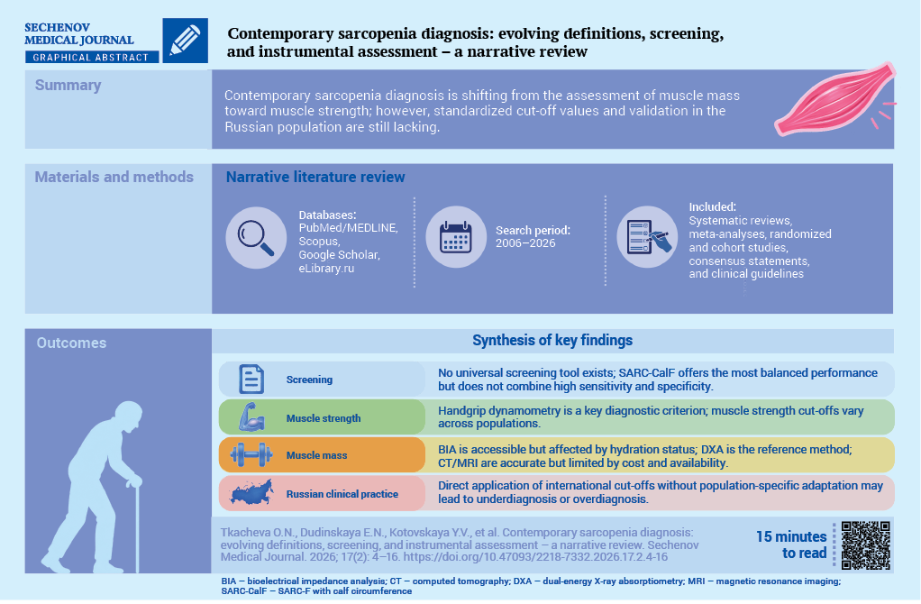
Sarcopenia currently affects 10–27% of people worldwide and 30–37% in the Russian Federation. Its social burden is considerable: the condition is associated with fractures, disability, and increased mortality. Sarcopenia nonetheless remains underdiagnosed. Diagnosis is hindered by the absence of universal criteria, standardized cutoff values, and widely available screening tools, while applying non-adapted foreign reference values to the Russian population may cause both under- and overdiagnosis.
Aim. To analyze the evolution of international and Russian approaches to sarcopenia diagnosis, assess the diagnostic value and limitations of available screening and instrumental methods, and formulate proposals for optimizing diagnostic strategies in clinical practice in the Russian Federation.
Materials and methods. A literature review on sarcopenia diagnosis was conducted using PubMed/MEDLINE, Scopus, Google Scholar, and eLibrary.ru for 2006–2026. Systematic reviews, meta-analyses, randomized controlled trials and cohort studies, consensus documents, and clinical guidelines were included.
Results. A shift was observed from the isolated assessment of muscle mass toward diagnostic models in which muscle strength serves as the primary criterion and physical performance as an indicator of disease severity. Handgrip dynamometry cutoff values varied across populations. In particular, muscle strength in Russians older than 65 years was at the lower limit of European reference values. No screening instrument demonstrated both high sensitivity and high specificity. The most balanced instrument, SARC-CalF, has not been validated in the Russian population. Instrumental methods are highly accurate; however, their use is limited by high cost, limited availability, dependence on hydration status, and the lack of standardized protocols.
Conclusion. To improve the effectiveness of sarcopenia diagnosis in Russia, national reference values should be developed, diagnostic protocols standardized, and screening instruments validated.
The relevance of sarcopenia research is determined by its direct impact on disability, hospitalizations, and mortality. Sarcopenia increases the risk of falls, fractures, and loss of independence in activities of daily living and is also associated with cardiovascular and respiratory diseases, as well as cognitive impairment [1][2]. According to the United Nations, the proportion of people aged ≥65 years increased from 6% in 1990 to 9% in 2019 and is projected to reach 16% by 20501. The global prevalence of sarcopenia ranges from 10% to 27% [3], whereas in the Russian Federation it ranges from 30% to 37% [4][5].
The aim of this review is to analyze the evolution of international and Russian approaches to sarcopenia diagnosis, assess the diagnostic value of available screening and instrumental methods, identify their limitations, and formulate proposals for optimizing diagnostic strategies in real-world clinical practice in the Russian Federation.
For this narrative review, a literature search was conducted in the electronic databases PubMed/MEDLINE, Scopus, Google Scholar, and eLibrary.ru for the period from January 2006 to June 2026. The search strategy included combinations of the following terms in Russian and English: “sarcopenia”, “diagnosis”, “screening”, “muscle mass”, “muscle strength”, “physical performance”, “bioimpedance analysis”, “dual-energy X-ray absorptiometry”, “dynamometry”, “older adults”, and “frailty.”
The inclusion criteria comprised systematic reviews, meta-analyses, randomized controlled trials, large observational cohort studies, consensus documents, and clinical guidelines issued by the European Working Group on Sarcopenia in Older People (EWGSOP and EWGSOP2), Asian Working Group for Sarcopenia (AWGS), Global Leadership Initiative in Sarcopenia (GLIS), Foundation for the National Institutes of Health Sarcopenia Project (FNIH), Sarcopenia Definition and Outcomes Consortium (SDOC), and the Russian Association of Gerontologists and Geriatricians (RAGG), as well as publications addressing the diagnosis, epidemiology, or pathogenesis of sarcopenia. The exclusion criteria were clinical case reports, animal studies, and publications not related to the diagnosis of sarcopenia.
Data extraction was performed independently by five authors, with disagreements resolved by consensus. For each included source, the following information was recorded: authors, year of publication, study design, study population, methods used for sarcopenia diagnosis, main findings, and limitations.
The first step toward standardizing sarcopenia diagnosis was the EWGSOP consensus (2010), which defined sarcopenia as a syndrome characterized by progressive loss of skeletal muscle mass and strength [6]. In 2014, the FNIH, based on data from large cohort studies, proposed optimized cutoff values for muscle strength and muscle mass [7].
In 2016, sarcopenia received a separate M62.84 code in the International Classification of Diseases, Tenth Revision, Clinical Modification, used in the United States (ICD-10-CM); the code became effective on October 1, 2016, formally recognizing sarcopenia as a distinct disease entity rather than merely a syndrome [8]. This step not only emphasized the need for standardized diagnosis and treatment but also promoted convergence of diagnostic approaches at the global level. It should be noted that this refers specifically to the national clinical modification: in the core ICD-10 version of the World Health Organization, which is also used in the Russian Federation, there is no separate category for sarcopenia, while code M62.8 denotes “Other specified disorders of muscle.”
In 2018, EWGSOP2 defined sarcopenia as a progressive and generalized skeletal muscle disorder [2]. In 2019, AWGS proposed ethnically adapted cutoff values [9]. SDOC emphasized the need to distinguish sarcopenia from cachexia and malnutrition [10], while GLIS (2024) separated diagnostic parameters into components and outcomes, including impaired physical performance, falls, and disability [11]. In 2025, AWGS published an updated consensus, extending the age range to 50–64 years, simplifying the diagnostic algorithm, and abandoning the category of “severe sarcopenia” in favor of “probable sarcopenia” [12].
In 2026, the clinical guidelines of the Russian Association of Gerontologists and Geriatricians defined sarcopenia as a progressive, age-associated, generalized, potentially reversible skeletal muscle disease associated with a high risk of falls, fractures, disability, and death2.
Despite substantial progress in understanding the pathogenetic mechanisms and clinical consequences of sarcopenia, its diagnosis remains challenging. This is due to the lack of universal diagnostic criteria, standardized cutoff values, and widely available screening tools.
According to the EWGSOP2, AWGS, and FNIH consensus recommendations, diagnosis follows a three-step approach: identification of reduced muscle strength, confirmation of low muscle mass, and, at the final stage, assessment of physical performance.
However, differences in the cutoff values used complicate interpretation of the results and comparison of data across populations.
Another major challenge is the lack of universal standards and readily accessible tools for diagnosing sarcopenia. Accurate methods for assessing muscle mass, including dual-energy X-ray absorptiometry (DXA), computed tomography (CT), and magnetic resonance imaging (MRI), are limited in routine use by their cost and technical requirements. Screening tools such as SARC-F (strength, assistance in walking, rise from a chair, climb stairs, falls) have low sensitivity at early stages.
No universal screening test combines high sensitivity, specificity, and reproducibility [2][13]. The SARC-F questionnaire is the most widely used screening tool; a score of ≥4 points indicates a risk of sarcopenia [14]. However, its low sensitivity (21–61%, with a specificity of 86–91%) limits its use [15–20].
To improve diagnostic accuracy, several modifications incorporating anthropometric parameters have been developed. The most extensively studied is SARC-CalF (SARC-F with calf circumference), which includes measurement of calf circumference (cutoff values <34 cm in men and <33 cm in women; 10 additional points, with a total cutoff of ≥11) [21]. According to E.J. Kim et al. [22], the sensitivity of SARC-CalF is 53.3%, specificity is 87.3%, and the area under the ROC curve (AUC) is 0.78. Other modifications include SARC-F + MUAC (mid-upper arm circumference), SARC-F + EBM (elderly body mass) where age ≥75 years and body mass index (BMI) ≤21 kg/m², SARC-CalF + MUAC, and SARC-CalF + MUAC adjusted for BMI. Adding MUAC increases sensitivity while reducing specificity [23], whereas SARC-F + EBM improves identification of older patients with low body mass [24][25]. These combined versions require further validation [23][26].
Among non-questionnaire-based methods, the Ishii test, with cutoff values of ≥105 points for men and ≥120 points for women [27], showed the highest diagnostic accuracy in the study by A.B. de Lima et al. [23]: in men, sensitivity was 69.2%, specificity 97.1%, and AUC 0.831; in women, the corresponding values were 52.5%, 100%, and 0.762.
The MSRA (Mini Sarcopenia Risk Assessment) questionnaire, available in MSRA-7 and MSRA-5 versions, assesses age, hospitalizations, physical activity, nutritional status, and weight loss [28]. Cutoff values of ≤30 points for MSRA-7 and ≤45 points for MSRA-5 are associated with an increased risk of sarcopenia [28][29]. MSRA has high sensitivity (80%) but comparatively lower specificity (50–60%); these diagnostic characteristics have been confirmed in validation studies conducted in different
populations [28–31].
The choice of screening tool depends on the clinical setting and available resources. Amongst the Russian population, validation data for the tools discussed above remain limited: SARC-F has been studied mainly in selected disease-specific groups and showed a low diagnostic value [19], whereas data on SARC-CalF, the Ishii test, and MSRA are lacking. Moreover, the diagnostic accuracy of screening tools varies substantially across studies and populations [32], underscoring the need for their separate validation before widespread use in Russian clinical practice.
Table 1 presents a comparative overview of the main screening tools used to identify sarcopenia.
Table 1. Comparative characteristics of screening instruments for the detection of sarcopenia
|
Test |
Components |
Risk cutoff |
Advantages |
Limitations |
|
SARC-F [14] |
5 questions: muscle strength, walking, rising from a chair, climbing stairs, falls |
≥4 points |
Simple, rapid, and does not require equipment |
Low sensitivity (21–61%); depends on cognitive status |
|
SARC-CalF [21][22] |
SARC-F + CC |
≥11 points; CC <34 cm in men, <33 cm in women |
Higher diagnostic accuracy than SARC-F |
Sensitivity ~53%; affected by edema and obesity |
|
SARC-F + |
SARC-F + MUAC |
≥12 points |
Increases sensitivity by incorporating upper-arm anthropometry |
Reduced specificity; possible false-positive results |
|
SARC-F + EBM [24] |
SARC-F + age ≥75 years + BMI ≤21 kg/m² |
≥12 points |
Better identification of patients with low body mass |
Depends on accurate assessment of BMI and age |
|
SARC-CalF + |
SARC-F + CC + MUAC |
≥12 points |
Balance between sensitivity and specificity |
Moderate accuracy; limited external validation |
|
BMI-adjusted SARC-CalF + MUAC [23] |
SARC-F + CC + MUAC adjusted for BMI |
Not standardized |
Potential applicability in patients with excess body weight |
Requires further validation |
|
Ishii test [27] |
Age + handgrip strength + CC |
≥105 points in men; ≥120 points in women |
High diagnostic accuracy |
Requires a dynamometer and calculator; population-specific cutoffs need clarification |
|
MSRA 7/ |
Age, hospitalizations, physical activity, nutrition, weight loss |
≤30 / ≤45 points |
High sensitivity (~80%); takes nutritional and social factors into account |
Low specificity (50–60%); depends on the accuracy of patient responses |
Note: BMI – body mass index; CC – calf circumference; EBM – elderly body mass; MSRA – Mini Sarcopenia Risk Assessment; MUAC – mid-upper arm circumference; SARC-CalF – SARC-F with calf circumference; SARC-F – strength, assistance in walking, rise from a chair, climb stairs, falls.
Handgrip dynamometry is a simple, inexpensive, and non-invasive method for assessing muscle strength and is widely used in geriatrics, rehabilitation, and epidemiological research. Low muscle strength is associated with an increased risk of functional limitations, hospitalization, cardiovascular events, and mortality [33][34], which supports its inclusion in diagnostic algorithms for sarcopenia and frailty.
EWGSOP2 and AWGS 2025 identified low muscle strength as a central diagnostic parameter alongside muscle mass, with reduced strength regarded as a key early sign [2][12]. However, the use of handgrip dynamometry is limited by variability in cutoff values, anthropometric differences, heterogeneity of measurement protocols, and differences between dynamometers [2][35].
Historically, dynamometry has been developing since the nineteenth century, while the hydraulic Jamar dynamometer became a reference instrument in the 1950s [36][37]. Analysis of the literature showed that even at that time measurement results were influenced by hand position, elbow flexion angle, number of attempts, and type of device [35][37]. The standardized protocol of the American Society of Hand Therapists recommends measurements in a seated position, with the shoulders adducted and the elbows flexed at 90° [38]. V. Mathiowetz et al. [39] established early normative values and confirmed an age-related pattern, with peak strength around the age of 30 years followed by a gradual decline. L.P. Fried et al. (2001) [40] included weakness in the frailty phenotype, considering handgrip strength a marker of vulnerability.
EWGSOP 2010 incorporated handgrip dynamometry into the diagnosis of sarcopenia, whereas EWGSOP2 substantially increased its diagnostic importance by defining low muscle strength as the primary criterion: values <27 kg in men and <16 kg in women indicate probable sarcopenia [2][6]. AWGS 2019 proposed cutoff values of <28 kg for men and <18 kg for women in Asian populations [9]. AWGS 2025 requires concurrent reductions in muscle strength and muscle mass for the diagnosis of sarcopenia; the cutoff values are <28 and <18 kg for individuals aged ≥ 65 years and < 34 and < 20 kg for those aged 50–64 years, respectively [12].
There is no universal cutoff value for muscle strength. EWGSOP2 relies on the study by R.M. Dodds et al. (60,803 observations; a T-score approach using a value 2.5 standard deviations below peak strength) [41]. However, muscle strength depends on height, body mass, ethnicity, physical activity, and the measurement protocol. Historically, higher cutoff values have been used, including approximately 30 kg in men and 20 kg in women [42], as well as 37 kg in men and 21 kg in women for identifying the risk of mobility limitation [43], which highlights the dependence of cutoff values on the selected clinical outcome and population characteristics.
International differences are evident: in the PURE study (125,462 participants), handgrip strength was higher in Europe and North America and lower in South Asia and Africa [44]. A 2024 systematic review including 2.4 million adults from 69 countries showed that peak handgrip strength values were 49.7 kg in men and 29.7 kg in women aged 30–39 years, followed by a subsequent decline [45]. Comparison of Russian and Norwegian populations aged 40–69 years revealed persistent between-country differences in strength that could not be explained by protocol differences [46]. Russian data showed that muscle strength in individuals older than 65 years was at the lower end of European reference ranges; subsequently, Russian cutoff values of 24 kg for men and 17 kg for women were proposed, although the sample was limited to individuals up to 74 years of age [47][48]. At the same time, the 2026 clinical guidelines for Russian practice adopted the EWGSOP2 cutoff values, making their validation in representative Russian cohorts a priority.
BMI-adjusted cutoff values are not intended for the diagnosis of sarcopenia [40]. A 2007 study found that muscle strength in Taiwanese individuals was 25–27% lower than the “consolidated norms,” which led to the development of regional reference values [49].
Methodological challenges include variability in measurement protocols, including patient position, number of attempts, and choice of hand [35], as well as differences between dynamometers.
Thus, muscle strength is not an independent diagnostic criterion for sarcopenia. Cutoff values depend on the population, age, sex, and the clinical outcome being assessed. Dynamometry requires standardization of the device, measurement protocol, and cutoff values. Otherwise, methodological variability may compromise the reproducibility of results.
Bioelectrical impedance analysis (BIA) is an accessible method for assessing body composition based on the electrical resistance of tissues, allowing estimation of muscle mass, fat mass, and body water content. The main parameter used for diagnosing sarcopenia is the appendicular skeletal muscle mass index (ASMI; appendicular muscle mass / height²). According to AWGS 2019, the BIA cutoff values for ASMI are <7.0 kg/m2 in men and <5.7 kg/m² in women [9].
However, not all devices are capable of separately estimating muscle mass in the arms and legs, which limits calculation of ASMI. In such cases, alternative indices are used, including the skeletal muscle index (total muscle mass / height²) or lean mass index, although their cutoff values are less standardized and require population-specific validation.
BIA also provides the phase angle as a marker of soft-tissue quality, reflecting metabolic characteristics and hydration status. Higher values are assumed to indicate greater cell membrane integrity [50][51]. Results may be distorted in the presence of metal or silicone implants, cardiac pacemakers, and the use of certain medications. No universally accepted phase-angle cutoff values for sarcopenia diagnosis have been established, highlighting the need for further research.
DXA in whole-body mode is a key method for assessing muscle mass and is based on differential X-ray absorption by soft tissues. Particular importance is placed on the assessment of appendicular muscle mass (AMM; the sum of muscle mass in the limbs) and ASMI. EWGSOP2 defines low muscle mass as an ASMI <7.0 kg/m² in men and <5.5 kg/m² in women, corresponding to AMM values of ≤20 kg and ≤15 kg, respectively [2].
In obese patients, alternative indices are used, including AMM/BMI (<0.789 in men and <0.512 in women according to FNIH) or AMM/body weight, which correlate more closely with physical performance [7]. AWGS 2025 proposes age-specific ASMI cutoff values: <7.2 kg/m² in men and <5.5 kg/m² in women aged 50–64 years, and <7.0 kg/m² and <5.4 kg/m², respectively, in individuals aged ≥65 years [12].
A meta-analysis in professional athletes showed that BIA systematically overestimates lean mass compared with DXA, by an average of 2.78 kg. However, these findings cannot be directly extrapolated to the general population because of differences in body composition [52]. In addition, DXA cannot distinguish skeletal muscle tissue from other components of lean mass, such as fluid, glycogen, and connective tissue, which may lead to overestimation of muscle mass in the presence of edema. The method is also subject to projection-related errors, including tissue overlap, positioning artifacts, and the presence of implants. DXA provides primarily quantitative assessment and does not directly characterize muscle quality, architecture, or fatty infiltration, which limits its prognostic value.
Table 2 summarizes the key parameters of BIA, DXA, and imaging-based methods.
Table 2. Comparative characteristics of instrumental methods for muscle mass assessment
|
Method |
Main parameters and cutoff values |
Advantages |
Limitations |
Recommendations for use |
|
BIA |
ASMI: <7.0 kg/m² in men, <5.7 kg/m² in women (AWGS 2019) [9]; phase angle: <4.05° in men, <3.55° in women [51] |
Accessibility, portability, absence of ionizing radiation, and low cost |
Dependent on hydration status, unable to assess muscle quality, and limited accuracy in obesity |
For screening and initial assessment of muscle mass when DXA is unavailable; hydration status must be taken into account |
|
DXA |
ASMI: <7.0 kg/m² in men, <5.5 kg/m² in women (EWGSOP2) [2]; AMM/BMI: <0.789 in men, <0.512 in women (FNIH) [7] |
“Gold standard” for quantitative assessment of body composition; high reproducibility |
Projection-related distortions, inability to assess muscle quality, ionizing radiation, high cost, and limited availability |
Preferred method for diagnostic confirmation when available; reference method for quantitative assessment |
|
CT / MRI |
SMI at L3: <52.4 cm²/m² in men, <38.5 cm²/m² in women (CT) [57]; analogous protocols for MRI [58] |
Direct visualization; assessment of cross-sectional muscle area and fatty infiltration (myosteatosis); absence of projection-related distortions |
High cost, limited availability, radiation exposure with CT, long examination time, labor-intensive segmentation, and lack of standardized cutoff values |
For in-depth assessment, including muscle quality and myosteatosis, as well as for research purposes |
Note: AMM – appendicular muscle mass; ASMI – appendicular skeletal muscle mass index; AWGS – Asian Working Group for Sarcopenia; BIA – bioelectrical impedance analysis; BMI – body mass index; CT – computed tomography; DXA – dual-energy X-ray absorptiometry; EWGSOP2 – European Working Group on Sarcopenia in Older People 2; FNIH – Foundation for the National Institutes of Health Sarcopenia Project; L3 – third lumbar vertebra; MRI – magnetic resonance imaging; SMI – skeletal muscle index.
One of the fundamental studies that established reference values for muscle mass in European populations was the Rosetta Study, a body composition research project. Its findings were used by R.N. Baumgartner et al. (1998) [53], who developed and validated an anthropometric equation for predicting AMM, using DXA as the reference method. The equation demonstrated high accuracy (R² = 0.91; standard error of the estimate = 1.58 kg), allowing its use in epidemiological studies when instrumental methods were unavailable. An operational definition of sarcopenia was proposed based on the AMM/height² index, with sarcopenia defined as a value more than 2 standard deviations below the mean of a young reference population. The cutoff values were 7.26 kg/m² for men and 5.45 kg/m² for women [53]. This approach subsequently formed the basis for later consensus definitions, including EWGSOP and FNIH. The Health ABC Study confirmed the applicability of these reference values and contributed to the development of the EWGSOP 2010 criteria [54].
Asian studies that subsequently informed the AWGS 2014 consensus demonstrated poor agreement with the Baumgartner equation for identifying low muscle mass [55]. The limitations of height-adjusted muscle mass assessment were particularly evident in women, as the index did not adequately reflect age-related loss of muscle mass because of the substantial decline in height with age. A 2011 study in a Chinese population reported similar findings: the AMM/height² cutoff values for Chinese adults (5.85 kg/m² for men and 4.23 kg/m² for women) were substantially lower than those reported for the US population (7.26 and 5.45 kg/m², respectively), while the cutoff value for women was even lower than that reported in Hong Kong (4.82 kg/m²) [56]. These findings called into question the universality of this approach and highlighted the need for ethnicity-specific criteria in Asian populations.
CT and MRI are among the most reliable methods for assessing muscle mass because they provide three-dimensional measurements and avoid the projection-related distortions inherent to DXA. CT allows assessment of muscle density in Hounsfield units and segmentation of muscle tissue on axial images, distinguishing it from adipose and connective tissue and enabling calculation of the skeletal muscle index. The conventional approach involves measuring the cross-sectional muscle area at the level of the third lumbar vertebra and normalizing it to the patient’s height, which correlates with total body muscle mass [57][58]. The routine use of CT and MRI for screening is limited by high cost, radiation exposure with CT, and the labor-intensive nature of manual image segmentation.
In current diagnostic algorithms, physical performance tests are used to stratify disease severity rather than to replace muscle mass assessment. According to EWGSOP2, severe sarcopenia is diagnosed when reduced muscle strength, low muscle mass, and impaired physical performance are present simultaneously. The most commonly used tests are the Timed Up and Go test, the 4-m gait speed test, and the Short Physical Performance Battery (SPPB), which assess mobility, balance, endurance, and lower-extremity function [2][59].
A Timed Up and Go result of ≥20 s indicates poor physical performance and, when accompanied by reduced muscle strength and muscle mass, severe sarcopenia [2][60]. Diagnostic accuracy varies across populations: in hospitalized older patients, a cutoff of ≥10.85 s showed a sensitivity of 67.0%, specificity of 88.7%, and AUC of 0.80 [61][62]; in older women, the AUC was 0.703, while a cutoff of 9.8 s yielded a sensitivity of 89.8% and specificity of 41.7% [63]. The advantages of the test include rapid administration and integrated assessment, whereas its limitations include the influence of neurological, vestibular, and cognitive impairment and the absence of a universally accepted cutoff [64].
The 4-m gait speed test, with a cutoff of ≤0.8 m/s, indicates impaired physical performance [2][65]. According to a meta-analysis, each 0.1 m/s decrease in gait speed is associated with a 12% increase in mortality risk [66]. However, the test has limited diagnostic value for sarcopenia [64]. A five-repetition chair stand time >15 s is interpreted as reduced lower-extremity strength and may be used as an alternative to handgrip dynamometry [2][59]. In the study by L.A. da Costa Teixeira et al. [63], the test demonstrated moderate diagnostic accuracy (AUC <0.7). Its limitations include the influence of joint pain, chair height, and performance technique.
The SPPB includes three components: balance tests, 4-m gait speed, and the five-repetition chair stand test; each component is scored from 0 to 4 points, with a total score ranging from 0 to 12 [67]. A score of ≤8 points indicates poor physical performance and can be used to confirm severe sarcopenia. In the study by S. Phu et al. [68], the SPPB demonstrated moderate diagnostic performance (AUC 0.644–0.770); a cutoff of ≤8 points showed high sensitivity (82–100%) but low specificity (36–41%). The limitations of the SPPB include the relatively longer administration time and moderate specificity.
To advance toward personalized, evidence-based diagnosis of sarcopenia in Russia, a number of coordinated scientific and organizational measures are required:
Sarcopenia represents a major global challenge for healthcare systems in the context of population ageing. However, its effective detection and management require population-specific epidemiological and clinical data. Direct application of international criteria to Russian clinical practice without appropriate adaptation and validation may reduce diagnostic accuracy and increase the risk of unnecessary healthcare expenditures.
An integrated diagnostic algorithm is needed that combines accessible screening tools, standardized dynamometry, and stepwise instrumental confirmation. Integration of international experience with national research will facilitate the development of a sarcopenia diagnostic system consistent with the principles of evidence-based medicine and the needs of the Russian healthcare system.
Olga N. Tkacheva: conceptualization, manuscript editing, approval of the final version. Ekaterina N. Dudinskaya: conceptualization and design, analysis and interpretation of literature data, writing and editing of the manuscript. Yulia V. Kotovskaya, Anton V. Naumov, Natalia O. Khovasova: literature search and collection, analysis and interpretation of literature data, manuscript preparation. Kristina O. Chepygova, Bagzhat I. Isaeva: literature search and analysis. All authors approved the final version of the article.
Ethics statement. Ethics committee approval and informed consent were not required because this article is a narrative review of previously published studies and did not involve direct participation of humans or animals.
Conflict of interests. All authors declare no conflicts of interest.
Financing. The article was prepared within the framework of the state assignment “AI-based method for diagnosing sarcopenia and presarcopenia”, registration number 1025101700006-3.
Use of artificial intelligence. The AI-based tool Consensus (2026) was used for preliminary literature search and source systematization. All AI-generated outputs were reviewed and edited by the authors. AI was not used for text writing, conclusion formulation, or graphic creation. The authors are responsible for the manuscript content.
1. United Nations, Department of Economic and Social Affairs, Population Division. World Population Ageing 2019: Highlights. New York: United Nations; 2020. (ST/ESA/SER.A/444). https://digitallibrary.un.org/record/3907988/files/WorldPopulationAgeing2019-Report.pdf (access date: 26.07.2026).
2. Ministry of Health of the Russian Federation. Clinical Guidelines. Sarcopenia in Older and Very Old Patients (approved by the Ministry of Health of the Russian Federation, 2026). https://cr.minzdrav.gov.ru/preview-cr/1053_1/ (access date: 26.07.2026).
1. Newman A.B., Kupelian V., Visser M., et al. Strength, but not muscle mass, is associated with mortality in the health, aging and body composition study cohort. J Gerontol A Biol Sci Med Sci. 2006 Jan; 61(1): 72–77. https://doi.org/10.1093/gerona/61.1.72. PMID: 16456196
2. Cruz-Jentoft A.J., Bahat G., Bauer J., et al. Sarcopenia: revised European consensus on definition and diagnosis. Age Ageing. 2019 Jan; 48(1): 16–31. https://doi.org/10.1093/ageing/afy169. Erratum in: Age Ageing. 2019 Jul 1; 48(4): 601. https://doi.org/10.1093/ageing/afz046. PMID: 30312372
3. Petermann-Rocha F., Balntzi V., Gray S.R., et al. Global prevalence of sarcopenia and severe sarcopenia: a systematic review and meta-analysis. J Cachexia Sarcopenia Muscle. 2022 Feb; 13(1): 86–99. https://doi.org/10.1002/jcsm.12783. Epub 2021 Nov 23. PMID: 34816624. EDN: TTXHLL
4. Safonova Yu.A., Zotkin E.G. The frequency of sarcopenia in older age groups: evaluation of diagnostic criteria. Rheumatology Science and Practice. 2020; 58(2): 147–153 (In Russian). https://doi.org/10.14412/1995-4484-2020-147-153. EDN: SPYYUX
5. Bocharova K.A., Gerasimenko A.V., Zhaboeva S.L. Studying of prevalence of sarcopenia at patients in outpatient health care system. Modern problems of science and education. 2014; 6: 1149. EDN: TGQWIX
6. Cruz-Jentoft A.J., Baeyens J.P., Bauer J.M., et al. Sarcopenia: European consensus on definition and diagnosis: report of the European working group on sarcopenia in older people. Age Ageing. 2010 Jul; 39(4): 412–423. https://doi.org/10.1093/ageing/afq034. Epub 2010 Apr 13. PMID: 20392703
7. McLean R.R., Shardell M.D., Alley D.E., et al. Criteria for clinically relevant weakness and low lean mass and their longitudinal association with incident mobility impairment and mortality: The Foundation for the National Institutes of Health (FNIH) sarcopenia project. J Gerontol A Biol Sci Med Sci. 2014 May; 69(5): 576–583. https://doi.org/10.1093/gerona/glu012. PMID: 24737560
8. Cao L., Morley J.E. Sarcopenia is recognized as an independent condition by an international classification of disease, tenth revision, clinical modification (ICD-10-CM) code. J Am Med Dir Assoc. 2016 Aug; 17(8): 675–677. https://doi.org/10.1016/j.jamda.2016.06.001. PMID: 27470918
9. Chen L.K., Woo J., Assantachai P., et al. Asian working group for sarcopenia: 2019 consensus update on sarcopenia diagnosis and treatment. J Am Med Dir Assoc. 2020 Mar; 21(3): 300–307. e2. https://doi.org/10.1016/j.jamda.2019.12.012. Epub 2020 Feb 4. PMID: 32033882. EDN: BZZZWA
10. Bhasin S., Travison T.G., Manini T.M., et al. Sarcopenia definition: the position statements of the sarcopenia definition and outcomes consortium. J Am Geriatr Soc. 2020 Jul; 68(7): 1410–1418. https://doi.org/10.1111/jgs.16372. Epub 2020 Mar 9. PMID: 32150289. EDN: WQVAGI
11. Kirk B., Cawthon P.M., Arai H., et al. The conceptual definition of sarcopenia: delphi consensus from the global leadership initiative in sarcopenia (GLIS). Age Ageing. 2024 Mar; 53(3): afae052. https://doi.org/10.1093/ageing/afae052. PMID: 38520141. EDN: ZMUGAQ
12. Chen L.K., Hsiao F.Y., Akishita M., et al. A focus shift from sarcopenia to muscle health in the Asian working group for sarcopenia 2025 consensus update. Nat Aging. 2025 Nov; 5(11): 2164–2175. https://doi.org/10.1038/s43587-025-01004-y. Epub 2025 Nov 4. PMID: 41188603. EDN: VUIWLO
13. Dent E., Morley J.E., Cruz-Jentoft A.J., et al. International clinical practice guidelines for sarcopenia (ICFSR): screening, diagnosis and management. J Nutr Health Aging. 2018; 22(10): 1148–1161. https://doi.org/10.1007/s12603-018-1139-9. PMID: 30498820. EDN: TMQBEV
14. Malmstrom T.K., Morley J.E. SARC-F: a simple questionnaire to rapidly diagnose sarcopenia. J Am Med Dir Assoc. 2013 Aug; 14(8): 531–532. https://doi.org/10.1016/j.jamda.2013.05.018. Epub 2013 Jun 25. PMID: 23810110.
15. Nishikawa H., Asai A., Fukunishi S., et al. Screening tools for sarcopenia. In Vivo. 2021 Nov-Dec; 35(6): 3001–3009. https://doi.org/10.21873/invivo.12595. PMID: 34697131
16. Malmstrom T.K., Miller D.K., Simonsick E.M., et al. SARC-F: a symptom score to predict persons with sarcopenia at risk for poor functional outcomes. J Cachexia Sarcopenia Muscle. 2016 Mar; 7(1): 28–36. https://doi.org/10.1002/jcsm.12048. Epub 2015 Jul 7. PMID: 27066316
17. Lu J.L., Ding L.Y., Xu Q., et al. Screening accuracy of SARC-F for sarcopenia in the elderly: a diagnostic meta-analysis. J Nutr Health Aging. 2021; 25(2): 172–182. https://doi.org/10.1007/s12603-020-1471-8. PMID: 33491031. EDN: YJLGZC
18. Ida S., Kaneko R., Murata K. SARC-F for screening of sarcopenia among older adults: a meta-analysis of screening test accuracy. J Am Med Dir Assoc. 2018 Aug; 19(8): 685–689. https://doi.org/10.1016/j.jamda.2018.04.001. Epub 2018 May 31. PMID: 29778639
19. Toroptsova N.V., Dobrovolskaya O.V., Efremova A.O., Nikitinskaya O.A. Diagnostic value of the SARC-f questionnaire and muscle strength tests for the detection of sarcopenia in patients with rheumatoid arthritis. Rheumatology Science and Practice. 2020; 58(6): 678–682 (In Russian). https://doi.org/10.47360/1995-4484-2020-678-682. EDN: QGKYYP
20. Barbosa-Silva T.G., Menezes A.M.B., Bielemann R.M., et al. Enhancing SARC-F: improving sarcopenia screening in the clinical practice. J Am Med Dir Assoc. 2016 Dec; 17(12): 1136–1141. https://doi.org/10.1016/j.jamda.2016.08.004. Epub 2016 Sep 17. PMID: 27650212
21. Bahat G., Oren M.M., Yilmaz O., et al. Comparing SARC-F with SARC-CalF to screen sarcopenia in community living older adults. J Nutr Health Aging. 2018; 22(9): 1034–1038. https://doi.org/10.1007/s12603-018-1072-y. PMID: 30379299. EDN: MQKXYP
22. Kim E.J., Hong S., Kim D.Y., Son Y.J. Diagnostic accuracy of the SARC-CalF in community-dwelling older adults: a systematic review and meta-analysis. Gerontologist. 2025 Nov; 65(12): gnaf258. https://doi.org/10.1093/geront/gnaf258. PMID: 41270807. EDN: QRAZNS
23. de Lima A.B., Merchant R.A., Henriques-Neto D. Diagnostic accuracy of sarcopenia screening tools in low-income older adults in Amazonas, Brazil. Aging Clin Exp Res. 2025 Oct; 37(1): 307. https://doi.org/10.1007/s40520-025-03180-8. PMID: 41165917. EDN: JBWJUV
24. Kurita N., Wakita T., Kamitani T., et al. SARC-F validation and SARC-F+EBM derivation in musculoskeletal disease: the SPSS-OK study. J Nutr Health Aging. 2019; 23(8): 732–738. https://doi.org/10.1007/s12603-019-1222-x. PMID: 31560031. EDN: UJSZOG
25. Krzymińska-Siemaszko R., Deskur-Śmielecka E., KaluźniakSzymanowska A., et al. Comparison of diagnostic performance of SARC-F and its two modified versions (SARC-CalF and SARCF+EBM) in community-dwelling older adults from Poland. Clin Interv Aging. 2020 Apr; 15: 583–594. https://doi.org/10.2147/CIA.S250508. PMID: 32425513. EDN: UJSSFC
26. Nascimento T.G., Fayh A.P.T., Silva F.M., et al. SARC-F and six modified versions: prognostic role for prolonged hospital stay and 1-year mortality in older inpatients. BMC Geriatr. 2026 Jan; 26(1): 220. https://doi.org/10.1186/s12877-026-07012-w. PMID: 41572171. EDN: WIBBXN
27. Ishii S., Tanaka T., Shibasaki K., et al. Development of a simple screening test for sarcopenia in older adults. Geriatr Gerontol Int. 2014 Feb; 14 Suppl 1: 93–101. https://doi.org/10.1111/ggi.12197. PMID: 24450566
28. Rossi A.P., Micciolo R., Rubele S., et al.Assessing the risk of sarcopenia in the elderly: the mini sarcopenia risk assessment (MSRA) questionnaire. J Nutr Health Aging. 2017; 21(6): 743–749. https://doi.org/10.1007/s12603-017-0921-4. PMID: 28537342. EDN: WMXJMM
29. Krzymińska-Siemaszko R., Deskur-Śmielecka E., Styszyński A., Wieczorowska-Tobis K. Polish translation and validation of the mini sarcopenia risk assessment (MSRA) questionnaire to assess nutritional and non-nutritional risk factors of sarcopenia in older adults. Nutrients. 2021 Mar; 13(4): 1061. https://doi.org/10.3390/nu13041061. PMID: 33805185
30. Akarapornkrailert P., Muangpaisan W., Boonpeng A., Daengdee D. Validation of the Thai version of SARC-F, MSRA-7, and MSRA-5 questionnaires compared to AWGS 2019 and sarcopenia risks in older patients at a medical outpatient clinic. Osteoporos Sarcopenia. 2020 Dec; 6(4): 205–211. https://doi.org/10.1016/j.afos.2020.11.006. Epub 2020 Nov 26. PMID: 33426310. EDN: LODXYC
31. Yang M., Hu X., Xie L., et al. Validation of the Chinese version of the mini sarcopenia risk assessment questionnaire in communitydwelling older adults. Medicine (Baltimore). 2018 Sep; 97(37): e12426. https://doi.org/10.1097/MD.0000000000012426. PMID: 30213020
32. Huang L., Shu X., Ge N., et al. The accuracy of screening instruments for sarcopenia: a diagnostic systematic review and meta-analysis. Age Ageing. 2023 Aug; 52(8): afad152. https://doi.org/10.1093/ageing/afad152. PMID: 37596923. EDN: XFWZEV
33. Cooper R., Kuh D., Hardy R. Objectively measured physical capability levels and mortality: systematic review and meta-analysis. BMJ. 2010 Sep; 341: c4467. https://doi.org/10.1136/bmj.c4467. PMID: 20829298
34. Wu Y., Wang W., Liu T., Zhang D. Association of grip strength with risk of all-cause mortality, cardiovascular diseases, and cancer in community-dwelling populations: a meta-analysis of prospective cohort studies. J Am Med Dir Assoc. 2017 Jun; 18(6): 551.e17–551.e35. https://doi.org/10.1016/j.jamda.2017.03.011. PMID: 28549705
35. Roberts H.C., Denison H.J., Martin H.J., et al. A review of the measurement of grip strength in clinical and epidemiological studies: towards a standardised approach. Age Ageing. 2011 Jul; 40(4): 423–429. https://doi.org/10.1093/ageing/afr051. PMID: 21624928
36. Bohannon R.W., Peolsson A., Massy-Westropp N., et al. Reference values for adult grip strength measured with a Jamar dynamometer: a descriptive meta-analysis. Physiotherapy. 2006 Mar; 92(1): 11–15. https://doi.org/10.1016/j.physio.2005.05.003
37. Sousa-Santos A.R., Amaral T.F. Differences in handgrip strength protocols to identify sarcopenia and frailty – a systematic review. BMC Geriatr. 2017 Oct; 17(1): 238. https://doi.org/10.1186/s12877-017-0625-y. PMID: 29037155. EDN: YILXPS
38. MacDermid J., Solomon G., Valdes K., eds. Clinical assessment recommendations. 3rd ed. Mount Laurel, NJ: American Society of Hand Therapists; 2015. 80 p. ISBN: 978-0-69252-515-9
39. Mathiowetz V., Kashman N., Volland G., et al. Grip and pinch strength: normative data for adults. Arch Phys Med Rehabil. 1985 Feb; 66(2): 69–74. PMID: 3970660
40. Fried L.P., Tangen C.M., Walston J., et al. Frailty in older adults: evidence for a phenotype. J Gerontol A Biol Sci Med Sci. 2001 Mar; 56(3): 146–157. https://doi.org/10.1093/gerona/56.3.m146. PMID: 11253156
41. Dodds R.M., Syddall H.E., Cooper R., et al. Grip strength across the life course: normative data from twelve British studies. PLoS One. 2014 Dec; 9(12): e113637. https://doi.org/10.1371/journal.pone.0113637. PMID: 25474696
42. Lauretani F., Russo C.R., Bandinelli S., et al. Age-associated changes in skeletal muscles and their effect on mobility: an operational diagnosis of sarcopenia. J Appl Physiol (1985). 2003 Nov; 95(5): 1851–1860. https://doi.org/10.1152/japplphysiol.00246.2003. PMID: 14555665
43. Sallinen J., Stenholm S., Rantanen T., et al. Hand-grip strength cut points to screen older persons at risk for mobility limitation. J Am Geriatr Soc. 2010 Sep; 58(9): 1721–1726. https://doi.org/10.1111/j.1532-5415.2010.03035.x. PMID: 20863331
44. Leong D.P., Teo K.K., Rangarajan S., et al. Reference ranges of handgrip strength from 125,462 healthy adults in 21 countries: a prospective urban rural epidemiologic (PURE) study. J Cachexia Sarcopenia Muscle. 2016 Dec; 7(5): 535–546. https://doi.org/10.1002/jcsm.12112. Epub 2016 Apr 12. PMID: 27104109
45. Tomkinson G.R., Lang J.J., Rubín L., et al. International norms for adult handgrip strength: a systematic review of data on 2.4 million adults aged 20 to 100+ years from 69 countries and regions. J Sport Health Sci. 2025 Dec; 14: 101014. https://doi.org/10.1016/j.jshs.2024.101014. Epub 2024 Dec 6. PMID: 39647778. EDN: BSSCKI
46. Cooper R., Shkolnikov V.M., Kudryavtsev A.V., et al. Betweenstudy differences in grip strength: a comparison of Norwegian and Russian adults aged 40-69 years. J Cachexia Sarcopenia Muscle. 2021 Dec; 12(6): 2091–2100. https://doi.org/10.1002/jcsm.12816. Epub 2021 Oct 3. PMID: 34605224. EDN: DPAEFR
47. Turusheva A.V., Frolova E.V., Degryse J. Development of reference ranges of handgrip strength among healthy adults 65+ in Northwest Russia: a prospective population-based cohort Crystal study. Russian Family Doctor. 2017; 21(4): 29–35 (In Russian). https://doi.org/10.17816/RFD2017429-35. EDN: YMQKLK
48. Kapustina A.V., Shalnova S.A., Kutsenko V.A., et al. Assessment of muscle strength using handgrip test in a middle-aged and elderly Russian population and its association with health characteristics. Cardiovascular Therapy and Prevention. 2023; 22(8S): 3792 (In Russian). https://doi.org/10.15829/1728-8800-2023-3792. EDN: LSHNBA
49. Wu S.W., Wu S.F., Liang H.W., et al. Measuring factors affecting grip strength in a Taiwan Chinese population and a comparison with consolidated norms. Appl Ergon. 2009 Jul; 40(4): 811–815. https://doi.org/10.1016/j.apergo.2008.08.006. Epub 2008 Oct 23. PMID: 18947819
50. Uemura K., Doi T., Tsutsumimoto K., et al. Predictivity of bioimpedance phase angle for incident disability in older adults. J Cachexia Sarcopenia Muscle. 2020 Feb; 11(1): 46–54. https://doi.org/10.1002/jcsm.12492. Epub 2019 Aug 22. PMID: 31436391
51. Yamada M., Kimura Y., Ishiyama D., et al. Phase angle is a useful indicator for muscle function in older adults. J Nutr Health Aging. 2019; 23(3): 251–255. https://doi.org/10.1007/s12603-018-1151-0. PMID: 30820513. EDN: EEYAOI
52. Dzator S., Weerasekara I., Shields M., et al. Agreement between dual-energy X-ray absorptiometry and bioelectric impedance analysis for assessing body composition in athletes: a systematic review and meta-analysis. Clin J Sport Med. 2023 Sep; 33(5): 557–568. https://doi.org/10.1097/JSM.0000000000001136. Epub 2023 Feb 28. PMID: 36853902. EDN: PLRBZJ
53. Baumgartner R.N., Koehler K.M., Gallagher D., et al. Epidemiology of sarcopenia among the elderly in New Mexico. Am J Epidemiol. 1998 Apr; 147(8): 755–763. https://doi.org/10.1093/oxfordjournals.aje.a009520. Erratum in: Am J Epidemiol 1999 Jun 15; 149(12): 1161. PMID: 9554417. EDN: IKMFOZ
54. Newman A.B., Kupelian V., Visser M., et al. Sarcopenia: alternative definitions and associations with lower extremity function. J Am Geriatr Soc. 2003 Nov; 51(11): 1602–1609. https://doi.org/10.1046/j.1532-5415.2003.51534.x. PMID: 14687390. EDN: EUBPTN
55. Kim Y.S., Lee Y., Chung Y.S., et al. Prevalence of sarcopenia and sarcopenic obesity in the Korean population based on the Fourth Korean National Health and Nutritional Examination Surveys. J Gerontol A Biol Sci Med Sci. 2012 Oct; 67(10): 1107–1113. https://doi.org/10.1093/gerona/gls071. Epub 2012 Mar 19. PMID: 22431554
56. Wen X., Wang M., Jiang C.M., Zhang Y.M. Are current definitions of sarcopenia applicable for older Chinese adults? J Nutr Health Aging. 2011 Dec; 15(10): 847–851. https://doi.org/10.1007/s12603-011-0088-3. PMID: 22159771. EDN: LQTHMO
57. Prado C.M., Lieffers J.R., McCargar L.J., et al. Prevalence and clinical implications of sarcopenic obesity in patients with solid tumours of the respiratory and gastrointestinal tracts: a population-based study. Lancet Oncol. 2008 Jul; 9(7): 629–635. https://doi.org/10.1016/S1470-2045(08)70153-0. Epub 2008 Jun 6. PMID: 18539529
58. Yao R., Gao B.L., Hu J., et al. Characteristics of mDIXON-Quant and proton magnetic resonance spectroscopy imaging of thigh skeletal muscles in patients with stroke-related sarcopenia: a prospective case-control study. J Yeungnam Med Sci. 2025; 42: 49. https://doi.org/10.12701/jyms.2025.42.49. Epub 2025 Aug 28. PMID: 40874399. EDN: DZKTAE
59. Ryu J.Y., Kim M., Kim K.S., et al. Chair stand test as a proxy for physical performance and muscle strength in sarcopenia diagnosis: the Korean frailty and aging cohort study. Aging Clin Exp Res. 2022 Oct; 34(10): 2449–2456. https://doi.org/10.1007/s40520-022-02172-2. Epub 2022 Aug 2. PMID: 35918606. EDN: EVLXBY
60. Podsiadlo D., Richardson S. The timed “Up & Go”: a test of basic functional mobility for frail elderly persons. J Am Geriatr Soc. 1991 Feb; 39(2): 142–148. https://doi.org/10.1111/j.1532-5415.1991.tb01616.x. PMID: 37050485
61. Martinez B.P., Gomes I.B., Oliveira C.S., et al. Accuracy of the Timed Up and Go test for predicting sarcopenia in elderly hospitalized patients. Clinics (Sao Paulo). 2015 May; 70(5): 369–372. https://doi.org/10.6061/clinics/2015(05)11. Epub 2015 May 1. PMID: 26039955
62. Pérez-Santiago L., Garzón-Hernández L.P., Martín-Arévalo J., et al. Accuracy of the Timed Up and Go Test for predicting low muscle mass in a preoperative prehabilitation program for colorectal cancer. J Clin Med. 2025 Mar; 14(6): 2088. https://doi.org/10.3390/jcm14062088. PMID: 40142896. EDN: LBUOVU
63. da Costa Teixeira L.A., Soares L.A., de Moura Oliveira M.C., et al. Diagnostic accuracy of sit-to-stand chair test and timed up and go test with proposed cutoff point for identifying sarcopenia in community-dwelling older women: a cross-sectional study. BMC Musculoskelet Disord. 2026 Jan; 27(1): 111. https://doi.org/10.1186/s12891-025-09470-6. PMID: 41507879. EDN: NJZGTQ
64. Looijaard S.M.L.M., Oudbier S.J., Reijnierse E.M. Single physical performance measures cannot identify geriatric outpatients with sarcopenia. J Frailty Aging. 2018; 7(4): 262–267. https://doi.org/10.14283/jfa.2018.19. PMID: 30298176
65. Tkacheva O.N., Kotovskaya Yu.V., Runikhina N.K., et al. Clinical Guidelines on Frailty. Russian Journal of Geriatric Medicine. 2025; 1(21): 6–48 (In Russian). https://doi.org/10.37586/2686-8636-1-2025-6-48. EDN: JCMOSK
66. Veronese N., Stubbs B., Volpato S., et al. Association between gait speed with mortality, cardiovascular disease and cancer: a systematic review and meta-analysis of prospective cohort studies. J Am Med Dir Assoc. 2018 Nov; 19(11): 981–988.e7. https://doi.org/10.1016/j.jamda.2018.06.007. Epub 2018 Jul 25. PMID: 30056008. EDN: BWXUVN
67. Guralnik J.M., Simonsick E.M., Ferrucci L., et al. A short physical performance battery assessing lower extremity function: association with self-reported disability and prediction of mortality and nursing home admission. J Gerontol. 1994 Mar; 49(2): 85–94. https://doi.org/10.1093/geronj/49.2.M85. PMID: 8126356. EDN: HJAWDD
68. Phu S., Kirk B., Bani Hassan E., et al. The diagnostic value of the Short Physical Performance Battery for sarcopenia. BMC Geriatrics. 2020 Jul; 20(1): 242. https://doi.org/10.1186/s12877-020-01642-4. PMID: 32660438. EDN: FWMQPA
Olga N. Tkacheva, Dr. of Sci. (Medicine), professor, corresponding member of the RAS, head of the Department of Diseases of Aging, Institute of Continuing Professional Education, director
16, 1st Leonova str., Moscow, 129226
Ekaterina N. Dudinskaya, Dr. of Sci. (Medicine), professor, Department of Diseases of Aging, Institute of Continuing Professional Education and Development, head of the Laboratory of Age-Related Metabolic and Endocrine Disorders, endocrinologist
16, 1st Leonova str., Moscow, 129226
Yulia V. Kotovskaya, Dr. of Sci. (Medicine), professor, deputy director for research, cardiologist of the highest qualification category
16, 1st Leonova str., Moscow, 129226
Anton V. Naumov, Dr. of Sci. (Medicine), professor, Department of Diseases of Aging, Institute of Continuing Professional Education and Development, head of the Laboratory of Musculoskeletal Disorders
16, 1st Leonova str., Moscow, 129226
Natalia O. Khovasova, Dr. of Sci. (Medicine), professor, Department of Diseases of Aging, Institute of Continuing Professional Education and Development, geriatrician, internist, senior researcher, Laboratory of Musculoskeletal Disorders
16, 1st Leonova str., Moscow, 129226
Bagzhat I. Isaeva, research physician
16, 1st Leonova str., Moscow, 129226
Kristina O. Chepygova, junior researcher, endocrinologist
16, 1st Leonova str., Moscow, 129226

|
1. Scale for the Assessment of Narrative Review Articles – SANRA | |
| Subject | ||
| Type | Исследовательские инструменты | |
Download
(246KB)
|
Indexing metadata ▾ | |
Sechenov Medical Journal. Editor's checklist for this article you can find here.
Журнал «Сеченовский вестник» |
| Sechenov Medical Journal |
Рецензии на рукопись |
| Peer-review reports |
Название / Title | Современная диагностика саркопении: эволюция определений, скрининг и инструментальная оценка – нарративный обзор / Contemporary sarcopenia diagnosis: evolving definitions, screening, and instrumental assessment – a narrative review |
Раздел / Section
| ВНУТРЕННИЕ БОЛЕЗНИ / INTERNAL DISEASES
|
Тип / Article | Обзор / Review |
Номер / Number | 1544
|
Страна/территория / Country/Territory of origin | Россия / Russia |
Язык / Language | Русский / Russian Английский / English
|
Источник / Manuscript source | Инициативная рукопись / Unsolicited manuscript |
Дата поступления / Received | 30.06.2026 |
Тип рецензирования / Type ofpeer-review | Двойное слепое / Double blind |
Язык рецензирования / Peer-review language | Русский / Russian
|
РЕЦЕНЗЕНТ А / REVIEWER A
Инициалы / Initials | 1544_А
|
Научная степень / Scientific degree | Кандидат медицинских наук / Candidate of Sci. (Medicine) |
Страна/территория / Country/Territory | Россия / Russia
|
Дата рецензирования / Date of peer-review | 01.09.2026 |
Число раундов рецензирования / Number of peer-review rounds | 2 |
Финальное решение / Final decision | принять к публикации / accept
|
ПЕРВЫЙ РАУНД РЕЦЕНЗИРОВАНИЯ / FIRST ROUND OF PEER-REVIEW
Обзор посвящен актуальной для современного медицинского сообщества теме саркопении, учитывая увеличение продолжительности жизни в РФ.
Статья написана очень подробно с освещением основных положений современных консенсусных документов и описанием методов диагностики саркопении с указанием тех особенностей, которые характерны для каждого существующего метода, что подчеркивает ее практическую значимость.
Важно отметить, что авторы указывают на необходимость разработки современных стандартов и нормативных показателей для российской популяции, что, несомненно, актуально для современной гериатрической практики.
Однако есть несколько замечаний:
ЗАКЛЮЧЕНИЕ: необходима доработка
ВТОРОЙ РАУНД РЕЦЕНЗИРОВАНИЯ / SECOND ROUND OF PEER-REVIEW
В представленном варианте авторы в полной мере учли все замечания и внесли изменения в соответствии с рекомендациями.
ЗАКЛЮЧЕНИЕ: принять к публикации
РЕЦЕНЗЕНТ B / REVIEWER B
Инициалы / Initials | 1544_В
|
Научная степень / Scientific degree | Кандидат медицинских наук / Candidate of Sci. (Medicine) |
Страна/территория / Country/Territory | Россия / Russia
|
Дата рецензирования / Date of peer-review | 29.08.2026
|
Число раундов рецензирования / Number of peer-review rounds | 2 |
Финальное решение / Final decision | принять к публикации / accept
|
ПЕРВЫЙ РАУНД РЕЦЕНЗИРОВАНИЯ / FIRST ROUND OF PEER-REVIEW
населения, ростом распространенности ожирения, высокой частотой встречаемости коморбидных пациентов в клинической практике. Отсутствие стандартных подходов к диагностике саркопении на территории Российской Федерации влечет за собой методологические ошибки при постановке диагноза, способные повлиять на тактику ведения пациента и прогноз.
Статья представляет собой обзор литературы, изложена на 21 странице, имеет 67 ссылок, 28 из которых были опубликованы за последние 6 лет. В статье приводятся 2 таблицы.
Статья оставляет приятное впечатление, следует глубокое понимание авторами изучаемой проблемы, тщательный анализ литературы, практическую направленность обзора литературы.
К статье есть несколько замечаний:
ЗАКЛЮЧЕНИЕ: необходима доработка и повторное рецензирование.
ВТОРОЙ РАУНД РЕЦЕНЗИРОВАНИЯ / SECOND ROUND OF PEER-REVIEW
Авторы тщательно переработали рукопись, благодаря чему ее качество значительно улучшилось. Все замечания были учтены.
Однако в связи с добавлением новых фрагментов текста возникли следующие замечания, которые могут быть учтены авторами совместно с редактором на этапе подготовки рукописи к публикации:
1) При прочтении исправленного вариант рукописи, в частности добавленного раздела «Материал и методы», возникает вопрос: в названии обзора (по крайней мере, на английском языке) указано, что он описательный (narrative). Не противоречит ли этому последний абзац вновь добавленного раздела?
2) Просьба к авторам и редактору на последующих этапах работы с рукописью еще раз внимательно прочитать весь текст, поскольку в рукописи, в том числе в исправленных фрагментах, все еще встречаются предложения/фрагменты, которые требуют правки, например:
«У мужчин чувствительность теста составила 69,2%, специфичность 97,1%, AUC 0,831; у женщин чувствительность теста составила 52,5%, 100% и 0,762 соответственно.»
ЗАКЛЮЧЕНИЕ: принять к публикации
РЕЦЕНЗЕНТ C / REVIEWER C
Инициалы / Initials | 1544_В
|
Научная степень / Scientific degree | Доктор медицинских наук / Doctor of Sci. (Medicine) |
Страна/территория / Country/Territory | Россия / Russia
|
Дата рецензирования / Date of peer-review | 04.09.2026
|
Число раундов рецензирования / Number of peer-review rounds | 2 |
Финальное решение / Final decision | принять к публикации / accept
|
ПЕРВЫЙ РАУНД РЕЦЕНЗИРОВАНИЯ / FIRST ROUND OF PEER-REVIEW
Название рукописи соответствует ее содержанию. Статья крайне актуальна и ведет к разработке отечественных пороговых значений для каждого диагностического компонента саркопении. Работа написана хорошим, литературным языком. Текст рукописи соответствует грамматическим нормам русского языка. Принципиальных замечаний нет, за исключением пожеланий, указанных в тексте рукописи.
Пожелания:
В связи с выходом КР «Саркопения у лиц пожилого и старческого возраста» рекомендуется употреблять термины, связанные с диагностикой саркопении, так, как они указаны в КР, иначе это создает путаницу в первую очередь для тех, кто будет применять диагностику саркопении на практике и в научных исследованиях.
В описании БИА следует указать ограничения этого метода: все виды имплантов, в том числе силиконовые, прием некоторых лекарственных препаратов.
Остается не понятным Ваш выбор в качестве скрининга опросника SARC CalF, чувствительность которого 53,3%, специфичность — 87,3% и практически не отличается от таковых показателей опросника SARC-F (21-61% и 86-91% соответственно). Они оба имеют низкую диагностическую точность. Поясните пожалуйста.
Расположите пожалуйста вначале содержание краткой батареи тестов физического функционирования (КТБФФ), чтобы было понятно, что тест ходьбы на 4 метра является его компонентом, а не отдельным функциональным тестом, а затем описание каждого из них.
ЗАКЛЮЧЕНИЕ: необходима доработка
ВТОРОЙ РАУНД РЕЦЕНЗИРОВАНИЯ / SECOND ROUND OF PEER-REVIEW
Авторы проделали огромную работу по исправлению замечаний рецензентов, проработав дополнительные литературные источники.
ЗАКЛЮЧЕНИЕ: принять к публикации
РЕКОМЕНДАЦИИ НАУЧНЫХ РЕДАКТОРОВ ЖУРНАЛА / RECOMMENDATIONS OF THE SCIENTIFIC EDITORS OF THE JOURNAL
 Email this article (Login required)
Email the author (Login required)
Email this article (Login required)
Email the author (Login required) 8-2 Trubetskaya st., Moscow, 119048
Federal State Autonomous Educational Institution of Нigher Education I.M.Sechenov First Moscow State Medical University of the Ministry of Health of the Russian Federation (Sechenovskiy University)
E-mail: sechenovmedj@staff.sechenov.ru
Processing of personal data





