



Contents
Scroll to:
https://doi.org/10.47093/2218-7332.2026.17.2.1489
Scroll to:
The neural plexus located on the ependyma’s apical surface of the cerebral ventricles may be an important link in the extra-barrier signal transmission and regulation of the cerebrospinal fluid composition, but to date it remains a poorly understood part of the central nervous system.
Aim. To investigate signs of functional activity of the supraependymal nerve plexus in early postnatal development using immunohistochemical methods.
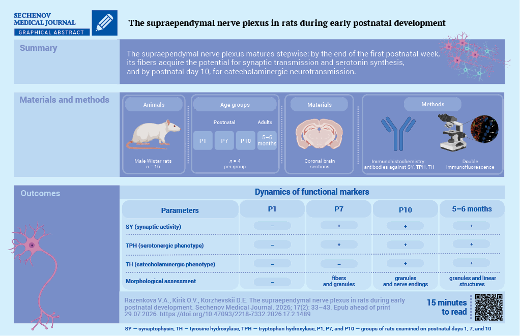
Materials and methods. Coronal brain sections of adult rats and rats on postnatal days 1, 7, and 10 (n = 16) were used for immunohistochemical analysis. Antibodies to tyrosine hydroxylase, tryptophan hydroxylase, and synaptophysin were used to identify supraependymal fibers and structures exhibiting potential synaptic activity.
Results. There was no immunopositive reaction observed in the supraependymal plexus of neonatal rats regardless of the marker used. Synaptophysin-containing puncta or fibers with varicosities on the surface of ependymal cells were first detected by the end of the first week of postnatal development. Tryptophan hydroxylase-positive structures were also observed at this stage of postnatal development. Fibers and puncta were located throughout the walls of the lateral and third ventricles, with the exception of the infundibular recess. Only on the 10th day of postnatal development were tyrosine hydroxylase-containing granules detected on the apical surface of the ependyma. A double immunofluorescence assay demonstrated that the tryptophan hydroxylase distribution matched with synaptophysin. Moreover, at none of the studied periods were neurons found on the surface of the ependyma.
Conclusion. The supraependymal plexus exhibits the capacity for synaptic transmission and serotonin synthesis by the end of the first week of postnatal development. By the 10th day of postnatal development, supraependymal nerve fibers may participate in catecholaminergic neurotransmission.
The biochemical composition of cerebrospinal fluid (CSF) serves as an important diagnostic criterion for a number of diseases, including meningitis, stroke, and brain abscess. The supraependymal nerve plexus is thought to be directly involved in regulating the composition and concentrations of biologically active substances in the CSF. The fibers forming this plexus are believed to be processes of neurons located in central nervous system nuclei that pass through the ependymal layer. It is also believed that these fibers are formed by processes of supraependymal neurons [1]. However, their presence has not yet been proven. The supraependymal plexus can be observed on the ependymal surface of the brain ventricular system: the lateral ventricles, the foramen of Monro, the third ventricle and the Sylvian aqueduct, as well as the floor of the fourth ventricle [2]. The apical surfaces of the cells covering the circumventricular organs constitute an exception [3].
Although supraependymal nerve fibers were described in the human brain in the 1980s [2][4], this component of the nervous system remains poorly understood. This is due to a combination of factors, including its specific location and the difficulty of identifying the supraependymal plexus in histological specimens. However, morphological studies in primates and other laboratory animals indicate the continuity of this structure across the mammalian phylogenetic lineage [2][5][6].
Another challenge in studying the supraependymal plexus is that most data concerning this structure were obtained in the 1970s and 1980s and therefore require careful verification and systematization. Currently, it is assumed that the supraependymal plexus is formed by fibers of serotonergic and catecholaminergic neurons [2][6–8]. It is also reported that some fibers and putative supraependymal neurons of the fourth ventricle contain glutamate and, therefore, may be glutamatergic [9]. Despite this, the neurotransmitter phenotype of the supraependymal fibers has not yet been fully established. Regarding their functional role, the main hypotheses can be summarized as follows: (1) the nerve fibers act as chemosensors of CSF composition; (2) they act on ependymal cells, influencing their secretory activity, shape, or ciliary activity; and (3) they release biologically active substances into the CSF that may act on cells of the choroid plexus and neurohypophysis, as well as on the circumventricular organs, including the area postrema, the subcommissural organ, and the subfornical organ [5]. In an attempt to assess the functional activity of the supraependymal plexus, it was shown that it possesses synaptic activity [7] and therefore may indeed secrete neurohormones or regulate the function of ependymal cells through asymmetric synapses. Nevertheless, none of these three hypotheses has yet been adequately tested experimentally.
Studies of the properties and functions of the supraependymal plexus in adult animals are numerous, but fragmentary, thus several questions about its development remain unclear. General knowledge of the signaling networks of the developing central nervous system suggests that supraependymal fibers may be involved in signal transmission as early as late embryonic or early postnatal development [10]. However, this question has not yet been definitively resolved. The developmental period during which supraependymal fibers acquire the ability to synthesize neurotransmitters also remains unknown.
The aim of this study was to investigate signs of functional activity of the supraependymal nerve plexus during early postnatal development using immunohistochemical methods.
The study material consisted of coronal brain sections obtained from male Wistar rats of four age groups: postnatal days 1, 7, and 10 (groups P1, P7, and P10, respectively) and adult animals aged 5–6 months (n = 16). The sample size was calculated using the resource equation method. There were four rats in each group. The animals were obtained from the Rappolovo breeding facility (Leningrad Oblast) and housed in a vivarium at room temperature under standard conditions, with free access to food and water. They were not subjected to any experimental interventions.
To obtain offspring and establish groups of animals at different stages of postnatal development, adult male Wistar rats were housed with females at a ratio of 1:3. Pregnancy was determined by the presence of sperm in vaginal smears. After insemination, the females were housed individually. After birth, the offspring were kept in cages with their mothers. Brain samples were collected on postnatal days 1, 7, and 10.
Animals were randomly selected for each stage of postnatal development (simple random sampling). Animals with obvious anatomical and/or behavioral abnormalities, as well as signs of distress, were excluded from the study. Before sampling, animals were euthanized by an overdose of ethyl ether vapor. Brain samples were fixed by immersion in a zinc-ethanol-formaldehyde solution and embedded in paraffin using standard methods. Sections 5 µm thick were cut from the paraffin blocks using a Leica RM 2125RT rotary microtome and mounted on HistoBond®+ adhesive-coated glass slides (Paul Marienfeld, Lauda-Königshofen, Germany). The specialist responsible for the collection, processing, and preparation of the biological material was not informed about the study design.
All series of histological sections were simultaneously processed using standardized immunohistochemical staining protocols. For the immunohistochemical detection of structures with potential synaptic activity, rabbit monoclonal antibodies (clone SJ26-85) against synaptophysin (SY; ET1606-56, Huabio, Hangzhou, China) were used at a dilution of 1:400.
To mark supraependymal serotonergic and catecholaminergic fibers, sheep polyclonal antibodies against tryptophan hydroxylase (TPH; 816401, BioLegend, San Diego, USA) at a dilution of 1:1000 and rabbit polyclonal antibodies against tyrosine hydroxylase (TH; ab112, Abcam, Cambridge, UK) at a dilution of 1:1000, respectively, were used.
For the negative control, one section from each processed series was incubated without primary antibodies; Antibody Diluent (Spring Bioscience, Pleasanton, USA) was applied instead of the primary antibody solution.
The IMMUNO View II detection system (1-1011-2, ImmunoRus, Moscow, Russia) and the VECTASTAIN Universal Quick HRP avidin-biotin kit (PK-8800, Vector Laboratories, Burlingame, USA) were used as secondary detection reagents. The immunohistochemical reaction product was visualized using 3,3'-diaminobenzidine chromogen included in the IMMUNO View II kit. Some sections were counterstained with alum hematoxylin. The resulting preparations were analyzed with a Leica DM750 microscope (Wetzlar, Germany) and photographed with a Leica ICC50 camera (Wetzlar, Germany).
To perform a double immunofluorescence reaction, antibodies to SY at a dilution of 1:100 and TPH at a dilution of 1:200 were mixed at a 1:1 ratio. A mixture of antibodies against goat and sheep immunoglobulins labeled with biotin (CTS008, R&D Systems, Minneapolis, USA) and Rhodamine Red X-conjugated antibodies against rabbit IgG (711-295-152, Jackson ImmunoResearch, Philadelphia, USA) were used as secondary reagents. After incubation in a mixture of secondary antibodies, sections were treated with a solution of Cy2-conjugated streptavidin (016-220-084, Jackson ImmunoResearch, Philadelphia, USA). The nuclear fluorescent dye Hoechst S769121 (3K010, Lumiprobe, Moscow, Russia) was used to stain cell nuclei.
The resulting preparations were analyzed using an LSM800 confocal laser scanning microscope with Airyscan system (Zeiss, Oberkochen, Germany). Fluorescence excitation was performed at 348 nm for Hoechst S769121, 488 nm for Cy2, and 561 nm for Rhodamine Red X. The resulting images were analyzed using ZEN3 software (Zeiss, Oberkochen, Germany). The specialist who analyzed the obtained histological preparations was not provided with any information about the study subjects.
In brain sections, SY immunoreactivity was detected throughout the tissue as round or oval granules. In neonatal rats, the reaction product could accumulate in the processes, forming a distinct pattern of nerve fiber bundles in the section. However, at this stage, no reaction to SY was observed on the apical surface of ependymal cells. SY-positive structures were first detected on the surface of ependymal cells in P7 rats as discretely arranged round granules or fibers with bead-like thickenings (Fig. 1). Fibers and granules were located along the walls of the lateral and third ventricles, except in the region of the infundibular recess. A similar distribution pattern of SY-positive structures was observed in P10 rats. However, SY immunoreactivity was no longer detected in fibers and was observed only as isolated granules on both the ependymal surface and within the nervous tissue. SY on the ependymal surface in adult animals was distributed similarly to that at P10.

FIG. 1. Distribution of synaptophysin, tryptophan hydroxylase, and tyrosine hydroxylase in the region of the foramen of Monro at different stages of postnatal development.
Notes: immunohistochemistry with nuclear counterstain by hematoxylin. Arrows indicate supraependymal structures; the asterisk indicates the cavity of the brain ventricles. Scale bar is 50 µm.
P1, P7, and P10 – denote groups of rats examined on postnatal days 1, 7, and 10, respectively.
TPH immunoreactivity revealed serotonin-producing neurons, their processes, and terminals in brain preparations from rats of all age groups studied. In P1 rats, no TPH immunoreactivity was detected on the ependymal surface. As with SY, round or oval TPH-positive granules appeared on the ependymal surface in P7 rats. TPH-immunopositive fibers running along the apical surface of ependymal cells were also detected in this group. In P10 rats, by contrast, extended structures were rarely detected, whereas TPH-immunopositive granules varied considerably in size, ranging from large granules approximately 6–8 μm in diameter to small granules 0.7–0.9 μm in diameter (Fig. 1). In adult animals, the TPH immunohistochemical reaction product accumulated in discrete, rounded granules arranged in chains on the ependymal surface.
Comparison of SY and TPH immunoreactivity in serial sections from the same animal showed that the distribution patterns of these marker proteins within the supraependymal nerve plexus overlapped. Like SY, TPH detected along the walls of the lateral ventricles and in the anterior regions, but not in the floor of the third ventricle.
TH immunohistochemistry revealed catecholamine-producing neurons and their processes along the entire length of the fibers. Interestingly, the TH immunohistochemical reaction product was absent from supraependymal elements not only in P1 rats but also in P7 rats (Fig. 1). TH-immunopositive granules on the apical surface of the ependyma were first detected in P10 rats. In adult animals, TH-immunopositive structures appeared as isolated granules and short linear structures with bead-like thickenings. The distribution of TH-immunopositive supraependymal structures corresponded to the localization of supraependymal fibers and synaptic terminals identified by SY and TPH immunohistochemistry.
A double immunofluorescence on brain sections from P7 and P10 rats and adult animals showed that most supraependymal plexus fibers were immunopositive for both markers (Fig. 2). Granules containing SY but not TPH were also observed. However, structures that may appear both immunopositive for TPH and immunonegative for SY were not observed in any of the cases studied.

FIG. 2. Synaptophysin (red) and tryptophan hydroxylase (green) in supraependymal plexus.
А. Region of the Sylvian aqueduct on postnatal day 7; scale bar is 20 µm.
B. Wall of the lateral ventricle on postnatal day 7, visualized using Airyscan technology; scale bar is 5 µm.
C. Anterior region of the third ventricle in an adult animal; three-dimensional reconstruction of a series of optical sections, Z-stack thickness, 4.5 µm; grid cell size, 20 × 20 µm; scale bar is 20 µm.
Note: double immunofluorescence staining with Hoechst S769121 nuclear counterstaining. The red channel indicates synaptophysin, and the green channel indicates tryptophan hydroxylase. Arrows indicate sites of potential synaptic activity containing synaptophysin; double arrows indicate structures containing both synaptophysin and tryptophan hydroxylase; the asterisk indicates the ventricular cavity.
The main objectives of this study were to trace the functional activity of the supraependymal nerve plexus, as well as to estimate the period at which the production of neurotransmitters begins.
To address the first objective, an immunohistochemical study was conducted using antibodies against SY, one of the proteins of the SNARE (soluble N-ethylmaleimide-sensitive factor attachment protein receptor) complex. SY acts as a regulator of neurotransmitter exocytosis from synaptic vesicles, so its appearance in synaptic terminals is traditionally considered a manifestation of synaptic activity by the cell [11]. During embryogenesis, SY begins to be detected at the 11th week of perinatal development in humans, and its appearance in certain areas of the brain coincides with the onset of synaptogenesis [12]. In the brain of laboratory rodents, SY appears in the perikarya of neurons by the end of the second week of embryonic development. However, detection of SY in nerve fibers and synaptic puncta is limited to day 17, and in some areas, even after birth, which also marks the onset of active synaptogenesis [13][14]. Our data show that despite the ubiquitous distribution of SY in the nervous tissue of the brain (Fig. 1), no positive immunohistochemical reaction is observed in the axons of the supraependymal nerve plexus on postnatal day 1. On postnatal day 7, clear SY immunoreactivity was detected not only in the presynaptic terminals but also along the plexus fibers, which may reflect their growth along the walls of the cerebral ventricles. By contrast, on postnatal day 10, SY was detected predominantly in nerve terminals and was absent from the fibers.
This SY distribution may be a consequence of the peculiarities of synaptogenesis in the supraependymal plexus. Neural connections formed during early development begin to organize complex networks by the end of the first postnatal week, and their formation is directly linked to critical periods of development. Thus, coordinated neuronal activity is first observed in the subcortical structures of the brain, which is associated with the need for motor coordination [15]. On postnatal day 9, after the sensory and motor functions development, a sharp increase in synaptogenesis in the cortex occurs [16], which is accompanied by the emergence of social behavior. Thus, the growth of axon cones on postnatal day 7 (as can be predicted from the results of the immunohistochemical reaction) and the formation of functional synapses on postnatal day 10 may indicate that the role of the supraependymal plexus may not be critical, at least during the first week of postnatal development.
To address the second objective, we compared the results of the immunohistochemical reaction to SY in consecutive sections of the same animal with the immunohistochemical reaction to TPH and TH, the enzymes that limit the synthesis of serotonin and catecholamines, respectively. Interest in these particular groups of neurotransmitters is due to previously accumulated data on the possible sources of fibers in the supraependymal nerve plexus [6][8], receptors present on the cells of the ventricular lining [17][18], and biologically active molecules released into the CSF via neurosecretion [19].
Indeed, analysis of the preparations showed that the fibers of the supraependymal plexus contained TPH as early as postnatal day 7 and both enzymes by postnatal day 10, indicating their potential ability to synthesize both serotonin and catecholamines. Serotonergic fibers are believed to originate from neurons concentrated in the raphe nuclei. Using anterograde (directed from neuron bodies to synaptic terminals) and retrograde (conversely, from neurites to neuron bodies) labeling, it was established that the fibers of the supraependymal nerve plexus in the lateral ventricle belong to cells of the dorsal and medial raphe nuclei [20]. Although some early studies suggested the presence of neuronal cell bodies capable of synthesizing serotonin within the ventricles themselves [21][22], there is currently no reason to believe there are serotonergic neurons located here. Analysis of the preparations obtained in the present study also revealed no structures that resembled neuronal cell bodies. However, the absence of neurons in the cerebral ventricles cannot be stated with complete certainty. While the sources of serotonergic axons have already been identified, the location of catecholaminergic nerve cells, whose fibers lie on the apical surface of the ependyma, remains unknown.
It is possible that the same neurons may be the source of these fibers. Mass spectrometric analysis performed in different brain regions revealed several nuclei containing cells synthesizing two classical neurotransmitters. In particular, in the dorsal raphe nucleus, one of the sources of supraependymal fibers, TPH colocalizes with norepinephrine in the same cells [23]. Further indirect confirmation of this hypothesis is provided by an earlier study conducted with transmission electron microscopy [20]. The authors demonstrated that a single synaptic terminal of a supraependymal axon can simultaneously contain not only electron-dense (i.e., serotoninergic) but also light-core vesicles. This suggests that TPH-containing axons can release co-transmitters. Thus, according to the results of the present study, these additional neurotransmitters may indeed be catecholamines. Moreover, it appears that catecholamine synthesis in axons of the supraependymal plexus begins later than serotonin synthesis.
The appearance of TH in fibers at a later developmental stage than TPH correlates with general concepts of the development of the catecholaminergic nervous system, the critical period of which is considered to be the first postnatal month [24][25]. In addition, it is known that serotonin is a biologically active molecule that influences the migration and positioning of neurons in the nervous tissue of the brain [26]. Also, serotonin in early postnatal development plays a role of the maturation and stabilization of glutamatergic dendritic spines [27], which indicates its involvement in the neural networks formation. These features are evolutionarily conserved in vertebrates, which ensure postnatal synaptogenesis, neurogenesis, and morphogenesis of the brain [26-28]. It may turn out that already formed serotonergic fibers of the supraependymal nerve plexus can form spatial and functional relationships with sprouting catecholaminergic axons.
Therefore, it cannot be ruled out that the catecholaminergic and serotonergic fibers of the supraependymal plexus may be structures that are distinct from one another. In this context, the result of a double immunofluorescence reaction for SY and TPH is of interest, demonstrating that these two proteins are not always colocalized in the same synaptic terminal. This means that other neurotransmitters could be released at such synaptic sites. These may include catecholamines, in particular norepinephrine [23], as well as other molecules, such as the already mentioned glutamate [9].
It is also worth mentioning the previously noted fact that the spatial distribution of supraependymal catecholaminergic fibers often coincides with the presence of the catecholaminergic subependymal plexus in the underlying nervous tissue [7]. Moreover, it is known that in certain areas of the brain, such as the dorsal striatum subventricular zone [8], the subfornical organ [29], and the nuclei of the mediobasal hypothalamus [18], TH-containing fibers can pass through the ependymal cell layer and come into contact with the CSF. Thus, one of these structures may be the source of TH-positive fibers.
This study was conducted on coronal brain sections which means the information content of the obtained data is limited. Therefore, the preparation and study of ventricular wall specimens appears to be a promising direction for further research.
The results of this study should be considered as pilot-stage findings due to the limited number of animals and the stages of postnatal development examined. Also, the data was obtained only from male rats. Thus, the observed timing of the appearance of functional activity markers remains approximate due to the lack of analysis of developmental stages other than the first, seventh, and tenth postnatal days. Due to the limited sample size, as well as the specific localization and detection of the study object in histological preparations, a quantitative assessment of immunopositive structures was not performed, making the study primarily descriptive. Therefore, the development of specialized methods for the morphometric characterization of the supraependymal nerve plexus is a pressing objective for future research.
This study allowed us to evaluate the dynamics of synaptic changes in the supraependymal plexus during early postnatal development. According to the results obtained, tryptophan hydroxylase and synaptophysin are present in a single fiber or nerve terminal at the seventh day of postnatal development. This suggests that supraependymal fibers are presumably capable of synthesizing and releasing monoamines by the end of the first week of postnatal development. The appearance of tyrosine hydroxylase in the studied structures is recorded later, on the tenth postnatal day. Further research is needed to clarify the co- or separate synthesis of serotonin and catecholamines by the same fibers of the supraependymal plexus, as well as the presence of neurons on the ependymal surface.
Valeria A. Razenkova: conducting the experiment, data collection, data analysis, drafting the manuscript. Olga V. Kirik: conducting the experiment, data analysis, drafting the manuscript. Dmitrii E. Korzhevskii: study concept and design, critical revision of the manuscript. All authors approved the final version of the article.
Ethics statements. The study was conducted in accordance with the European Convention for the Protection of Vertebrate Animals Used for Experimental and Other Scientific Purposes and was approved by the Local Ethics Committee of the Institute of Experimental Medicine (Protocol No. 1/25 dated January 23, 2025).
Data availability. The data supporting the findings of this study are available from the authors upon reasonable request.
Conflict of interest. The authors declare that there is no conflict of interest.
Financing. The study was supported by the State assignment of the Institute of Experimental Medicine, project no. FGWG-2025-0003.
Use of artificial intelligence. No artificial intelligence tools were used in the preparation of this manuscript.
Acknowledgments. The authors thank the staff of the Center for Collective Use of Scientific Equipment “Cellular and Molecular Technologies” of the Federal State Budgetary Scientific Institution “Institute of Experimental Medicine” (Saint Petersburg, Russia) for providing access to the equipment and for technical support.
1. Martínez P.M., de Weerd H. The fine structure of the ependymal surface of the recessus infundibularis in the rat. Anat Embryol (Berl). 1977 Dec; 151(3): 241–265. https://doi.org/10.1007/BF00318929. PMID: 603079
2. Richards J.G., Lorez H.P., Colombo V.E., et al. Demonstration of supra-ependymal 5-HT nerve fibres in human brain and their immunohistochemical identification in rat brain. J Physiol (Paris). 1981; 77(2–3): 219–224. PMID: 7026769
3. Rodríguez P., Bouchaud C. The supra-ependymal innervation is not responsible for the repression of tight junctions in the rat cerebral ependyma. Neurobiology (Bp). 1996; 4(3): 185–201. PMID: 9044345
4. Richards J.G., Lorez H.P., Colombo V.E., et al. Supraependymal nerve fibres in human brain: correlative transmission and scanning electron microscopical and fluorescence histochemical studies. Neuroscience. 1980; 5(8): 1489–1502. https://doi.org/10.1016/0306-4522(80)90011-1. PMID: 7402482
5. Lorez H.P., Richards J.G. Supra-ependymal serotoninergic nerves in mammalian brain: morphological, pharmacological and functional studies. Brain Res Bull. 1982 Jul-Dec; 9(1–6): 727–741. https://doi.org/10.1016/0361-9230(82)90179-4. PMID: 6184136
6. Michaloudi H.C., Papadopoulos G.C. Catecholaminergic and serotoninergic fibres innervate the ventricular system of the hedgehog CNS. J Anat. 1996 Oct; 189(Pt 2): 273–283. PMID: 8886949
7. Razenkova V.A., Kirik O.V. Synaptophysin expression by supraependymal structures of the rat brain. Cell Tiss. Biol. 2023; 65(4): 348–353 (In Russian). https://doi.org/10.31857/S0041377123040107. EDN: ZKYDBB
8. Troshev D., Bannikova A., Blokhin V., et al. Striatal neurons partially expressing a dopaminergic phenotype: functional significance and regulation. Int J Mol Sci. 2022 Sep; 23(19): 11054. https://doi.org/10.3390/ijms231911054. PMID: 36232359
9. Robinson S.R., Noone D.F., O’Dowd B.S. Ependymocytes and supra-ependymal axons in rat brain contain glutamate. Glia. 1996 Aug; 17(4): 345–348. https://doi.org/10.1002/(SICI)1098-1136(199608)17:4<345::AID-GLIA9>3.0.CO;2-Y. PMID: 8856331. EDN: AVNPVY
10. Hennou S., Khalilov I., Diabira D., et al. Early sequential formation of functional GABA(A) and glutamatergic synapses on CA1 interneurons of the rat foetal hippocampus. Eur J Neurosci. 2002 Jul; 16(2): 197–208. https://doi.org/10.1046/j.14609568.2002.02073.x. PMID: 12169102
11. Kolos Ye.A., Grigoriyev I.P., Korzhevskiy D.E. A synaptic marker synaptophysin. Morphology. 2015; 147(1): 78–82 (In Russian). EDN: TIJLST
12. Sarnat H.B., Born D.E. Synaptophysin immunocytochemistry with thermal intensification: a marker of terminal axonal maturation in the human fetal nervous system. Brain Dev. 1999 Jan; 21(1): 41–50. https://doi.org/10.1016/s0387-7604(98)00068-0. PMID: 10082252
13. Leclerc N., Beesley P.W., Brown I., et al. Synaptophysin expression during synaptogenesis in the rat cerebellar cortex. J Comp Neurol. 1989 Feb; 280(2): 197–212. https://doi.org/10.1002/cne.902800204. PMID: 2494237
14. Ovtscharoff W., Bergmann M., Marquèze-Pouey B., et al. Ontogeny of synaptophysin and synaptoporin in the central nervous system: differential expression in striatal neurons and their afferents during development. Brain Res Dev Brain Res. 1993 Apr; 72(2): 219–225. https://doi.org/10.1016/0165-3806(93)90186-e. PMID: 8485845
15. Dehorter N., Vinay L., Hammond C., Ben-Ari Y. Timing of developmental sequences in different brain structures: physiological and pathological implications. Eur J Neurosci. 2012 Jun; 35(12): 1846–1856. https://doi.org/10.1111/j.1460-9568.2012.08152.x. PMID: 22708595. EDN: PGKMRJ
16. Naskar S., Narducci R., Balzani E., et al. The development of synaptic transmission is time-locked to early social behaviors in rats. Nat Commun. 2019 Mar; 10(1): 1195. https://doi.org/10.1038/s41467-019-09156-3. PMID: 30867422. EDN: REVGHL
17. Li X., Wang S., Zhang D., et al. The periaxonal space as a conduit for cerebrospinal fluid flow to peripheral organs. Proc Natl Acad Sci USA. 2024 Nov; 121(45): e2400024121. https://doi.org/10.1073/pnas.2400024121. Epub 2024 Nov 1. PMID: 39485799. EDN: AQVPEZ
18. Meister B., Hökfelt T., Tsuruo Y., et al. DARPP-32, a dopamine- and cyclic AMP-regulated phosphoprotein in tanycytes of the mediobasal hypothalamus: distribution and relation to dopamine and luteinizing hormone-releasing hormone neurons and other glial elements. Neuroscience. 1988 Nov; 27(2): 607–622. https://doi.org/10.1016/0306-4522(88)90292-8. PMID: 2905789
19. Murtazina A.R., Bondarenko N.S., Pronina T.S., et al. A comparative analysis of CSF and the blood levels of monoamines as neurohormones in rats during ontogenesis. Acta Naturae. 2021 OctDec; 13(4): 89–97. https://doi.org/10.32607/actanaturae.11516. PMID: 35127152. EDN: VKGBIM
20. Tong C.K., Cebrián-Silla A., Paredes M.F., et al. Axons take a dive: specialized contacts of serotonergic axons with cells in the walls of the lateral ventricles in mice and humans. Neurogenesis (Austin). 2014 Jan; 1(1): e29341. https://doi.org/10.4161/neur.29341. PMID: 26413556
21. Cupédo R.N.J. The surface ultrastructure of the habenular complex of the rat. Anat Embryol (Berl). 1977 Dec; 152(1): 43–64. https://doi.org/10.1007/BF00341434. PMID: 605996
22. Cupédo R.N.J., de Weerd H. Serotonergic intraventricular axons in the habenular region. Phagocytosis after induced degeneration. Anat Embryol (Berl). 1980; 158(2): 213–226. https://doi.org/10.1007/BF00315907. PMID: 7356178
23. Sugiyama E., Guerrini M.M., Honda K., et al. Detection of a high-turnover serotonin circuit in the mouse brain using mass spectrometry imaging. iScience. 2019 Oct; 20: 359–372. https://doi.org/10.1016/j.isci.2019.09.036. Epub 2019 Sep 27. PMID: 31614319
24. Kalinina T.S., Dygalo N.N. Development of the noradrenergic system of the rat brain after prenatal exposure to corticosterone. Biology Bulletin. 2013; 40(6): 545–549. https://doi.org/10.1134/S1062359013040043. EDN: SKZMQN
25. Sukhareva E.V., Kalinina T.S., Bulygina V.V., Dygalo N.N. Tyrosine hydroxylase of the brain and its regulation by glucocorticoids. Vavilov Journal of Genetics and Breeding. 2016; 20(2): 212–219 (In Russian). https://doi.org/10.18699/VJ16.156. EDN: WLVJZL
26. García-González D., Khodosevich K., Watanabe Y., et al. Serotonergic projections govern postnatal neuroblast migration. Neuron. 2017 May; 94(3): 534–549.e9. https://doi.org/10.1016/j.neuron.2017.04.013. PMID: 28472655
27. Chang V.N., Ogelman R., Vargas R.S., Oh W.C. Serotonergic modulation of excitatory synapse development and plasticity. Mol Cells. 2026 May; 49(5): 100346. https://doi.org/10.1016/j.mocell.2026.100346. Epub 2026 Mar 10. PMID: 41819288. EDN: PXVANM
28. Udoh U.G., Bruno J.R., Osborn P.O., Pratt K.G. Serotonin strengthens a developing glutamatergic synapse through a PI3Kdependent mechanism. J Neurosci. 2024 Feb; 44(6): e1260232023. https://doi.org/10.1523/JNEUROSCI.1260-23.2023. PMID: 38169457. EDN: TKWRLG
29. Razenkova V.A., Korzhevskii D.E. Catecholaminergic structures of the rat subfornical organ. Cell and Tissue Biology. 2022; 16(6): 568–575. https://doi.org/10.1134/S1990519X22060062. EDN: NLALOY
Valeria A. Razenkova, Cand. of Sci. (Biology), Research Associate, Laboratory of functional morphology of the central and peripheral nervous system
12, Akademika Pavlova str., Saint Petersburg, 197022
Olga V. Kirik, Cand. of Sci. (Biology), Senior Researcher, Laboratory of functional morphology of the central and peripheral nervous system
12, Akademika Pavlova str., Saint Petersburg, 197022
Dmitrii E. Korzhevskii, Dr. of Sci. (Medicine), Professor of the RAS, Head of the Laboratory of functional morphology of the central and peripheral nervous system
12, Akademika Pavlova str., Saint Petersburg, 197022

|
1. The ARRIVE guidelines 2.0: author checklist | |
| Subject | ||
| Type | Исследовательские инструменты | |
Download
(107KB)
|
Indexing metadata ▾ | |
Журнал «Сеченовский вестник» |
| Sechenov Medical Journal |
Рецензии на рукопись |
| Peer-review reports |
Название / Title | Супраэпендимное нервное сплетение крыс в раннем постнатальном онтогенезе / The supraependymal nerve plexus in rats during early postnatal development |
Раздел / Section
| КЛЕТОЧНАЯ БИОЛОГИЯ / CELL BIOLOGY
|
Тип / Article | Оригинальная статья / Original article |
Номер / Number | 1489
|
Страна/территория / Country/Territory of origin | Россия / Russia |
Язык / Language | Русский / Russian Английский / English
|
Источник / Manuscript source | Инициативная рукопись / Unsolicited manuscript |
Дата поступления / Received | 16.04.2026
|
Тип рецензирования / Type ofpeer-review | Двойное слепое / Double blind |
Язык рецензирования / Peer-review language | Русский / Russian
|
РЕЦЕНЗЕНТ А / REVIEWER A
Инициалы / Initials | 1489_А
|
Научная степень / Scientific degree | Доктор медицинских наук / Doctor of Sci. (Medicine)
|
Страна/территория / Country/Territory | Россия / Russia
|
Дата рецензирования / Date of peer-review | 10.06.2026 |
Число раундов рецензирования / Number of peer-review rounds | 2 |
Финальное решение / Final decision | принять к публикации / accept
|
ПЕРВЫЙ РАУНД РЕЦЕНЗИРОВАНИЯ / FIRST ROUND OF PEER-REVIEW
Супраэпендимное нервное сплетение остается одной из наименее изученных периферических структур ЦНС, особенно в аспекте его постнатального развития; большинство имеющихся данных датируется работами 1970–80-х гг. и получено преимущественно на взрослых животных. Представленная работа восполняет существенный пробел, впервые описывая динамику становления данной структуры в критический период раннего постнатального онтогенеза, что имеет прямое отношение к проблемам раннего синаптогенеза, нейрорегуляции состава ликвора и перивентрикулярного нейрогенеза.
Авторами применен современный арсенал гистохимических подходов: традиционную иммуногистохимию с хромогенной визуализацией, двойную иммунофлуоресценцию и конфокальную микроскопию. Двойное мечение подтверждает высокую специфичность локализации маркеров и исключает артефакты наложения сигналов.
Научная новизна работы заключается в первом экспериментальном доказательстве гетерохронного становления функциональной активности супраэпендимного нервного сплетения: синаптическая компетентность и способность к синтезу серотонина формируются к 7-м постнатальным суткам, тогда как катехоламинергический компонент — только к 10-м суткам.
Работа выполнена на актуальную фундаментальную тему на высоком методическом уровне, прекрасно иллюстрирована. У рецензента нет замечаний по существу исследования, однако есть отдельные вопросы и пожелания по разделам «Введение» и «Обсуждение».
Использование авторами термина «супраэпендимные нейроны» является дискуссионным и требует существенных терминологических уточнений, поскольку в научной литературе отсутствует единый консенсус по этому вопросу. Авторы демонстрируют внутреннюю противоречивость в использовании данного термина. Так, во введении указано, что «… нервные волокна образованы супраэпендимными нейронами, а также клетками нервных центров, посылающих отростки сквозь эпендимный слой»; в то же время в обсуждении авторы сами признают, что существование истинных нейронов на поверхности эпендимы не доказано, однако продолжают использовать термин «супраэпендимные нейроны» без оговорок. У читателя-морфолога, не погруженного столь глубоко в проблему, может возникнуть впечатление, что на поверхности эпендимы повсеместно располагаются полноценные нейроны, тогда как преобладающий компонент сплетения — терминали аксонов, происходящих из отдаленных ядер.
В настоящей работе, выполненной с помощью достаточно специфических современных методов, наличие супраэпендимных нейронов также не выявлено; этот момент целесообразно специально зафиксировать не только в обсуждении, но и в аннотации и заключении.
Вопросы, касающиеся функций супраэпендимного сплетения (хемосенсорика, регуляция эпендимы, нейросекреция) не входят в число задач исследования, сама работа демонстрирует структурное созревание сплетений, но не предоставляет прямых доказательств в пользу какой-либо их функциональной роли. Поэтому эту новеллу более логично развернуть не во введении, а в разделе «Обсуждение».
ЗАКЛЮЧЕНИЕ: необходима доработка
ВТОРОЙ РАУНД РЕЦЕНЗИРОВАНИЯ / SECOND ROUND OF PEER-REVIEW
В переработанный вариант статьи авторами внесены необходимые уточнения и редакционные коррективы с учетом пожеланий рецензента. Работа может быть рекомендована к публикации.
ЗАКЛЮЧЕНИЕ: принять к публикации
РЕЦЕНЗЕНТ B / REVIEWER B
Инициалы / Initials | 1489_В
|
Научная степень / Scientific degree | Кандидат медицинских наук / Candidate of Sci. (Medicine)
|
Страна/территория / Country/Territory | Россия / Russian
|
Дата рецензирования / Date of peer-review | 12.06.2026 |
Число раундов рецензирования / Number of peer-review rounds | 2 |
Финальное решение / Final decision | принять к публикации / accept
|
ПЕРВЫЙ РАУНД РЕЦЕНЗИРОВАНИЯ / FIRST ROUND OF PEER-REVIEW
Представленная статья посвящена изучению супраэпендимного нервного сплетения крыс в раннем постнатальном онтогенезе с использованием иммуногистохимических методов. Работа направлена на оценку сроков появления маркеров синаптической активности и моноаминергической нейротрансмиссии в супраэпендимных структурах желудочковой системы головного мозга.
Актуальность исследования не вызывает сомнений. Супраэпендимное нервное сплетение остается недостаточно изученной структурой ЦНС, несмотря на его потенциальную роль в регуляции состава спинномозговой жидкости, взаимодействии с эпендимой и участии в нейросекреторных процессах. Особенно значимым является изучение этой структуры в раннем постнатальном периоде, поскольку именно в это время происходят интенсивные процессы формирования нейральных сетей, синаптогенеза и становления нейромедиаторных систем.
Научная новизна работы заключается в попытке определить сроки появления синаптофизин-, триптофангидроксилаза- и тирозингидроксилаза-иммунопозитивных структур в составе супраэпендимного сплетения у крыс на 1-е, 7-е и 10-е сутки постнатального развития. Интерес представляет вывод авторов о более раннем появлении признаков серотонинергической активности по сравнению с катехоламинергической. Дополнительную ценность работе придает использование двойной иммунофлуоресцентной реакции для сопоставления распределения синаптофизина и триптофангидроксилазы.
Иллюстративный материал достаточно высокого качества, в целом и полном соответствует содержанию статьи и демонстрирует основные результаты исследования. Компоновка панелей по принципу «маркер × возраст» удачна и позволяет визуально сопоставить динамику иммуногистохимической реакции. Подпись к рисунку содержит необходимые пояснения: указаны метод окрашивания, значение «стрелок и звездочек», возрастные группы и масштабный отрезок. В подписи желательно уточнить, что масштабный отрезок 50 мкм относится ко всем панелям рисунка.
Список литературы включает как классические работы 1970-1990-х годов, так и современные публикации. Наличие значительного числа ранних источников оправдано спецификой темы, поскольку многие фундаментальные данные о супраэпендимных волокнах были получены именно в этот период. Вместе с тем доля публикаций последних лет ограничена, поэтому список литературы желательно, при возможности, дополнить современными источниками по постнатальному развитию эпендимы, желудочковой зоны, серотонинергической регуляции, нейрогенным нишам и механизмам созревания синаптических контактов.
Основное замечание касается преимущественно описательного характера исследования. В работе отсутствует количественная оценка иммунопозитивных структур, количестве исследованных срезов и полей зрения, а также не описан статистический анализ. Это ограничивает доказательность выводов и делает часть заключений скорее качественными, чем строго количественно подтвержденными.
Желательно уточнить, проводился ли анализ препаратов вслепую и каким образом исключалась неспецифическая реакция.
Ряд формулировок в обсуждении и заключении следует сделать более «осторожными». Наличие синаптофизина и ферментов синтеза моноаминов указывает на потенциально функциональную активность структур, однако само по себе не доказывает факт нейромедиаторного высвобождения или полноценной синаптической передачи. Поэтому выводы о «синаптической активности» и «моноаминергической активности» желательно формулировать как морфологические признаки потенциальной функциональной зрелости.
Также, желательно усилить обсуждение ограничений исследования. Авторы указывают пилотный характер работы, малое количество животных, ограниченное число сроков и использование только самцов, однако эти ограничения следует связать с интерпретацией результатов. Особенно важно подчеркнуть, что промежуточные сроки между P1, P7 и P10 не исследовались, поэтому точный момент появления маркеров остается приблизительным.
В тексте встречаются отдельные стилистические и терминологические неточности, которые требуют редакторской правки. Согласно общим правилам орфографии, прилагательные, образованные от имён учёных в научных терминах, пишутся со строчной буквы. Поэтому, желательно ввести терминологические названия структур (межжелудочковое отверстие - отверстие монро). Ну и позволю себе замечание напоследок - термин «иммуноокрашивание» – не совсем корректен, это не окрашивание, это биохимическая реакция, несмотря на то что термин понемногу устаивается, предложу использовать: иимунопозитивная, иимунореактивная, ТГ-положительная структура.
ЗАКЛЮЧЕНИЕ: необходима доработка.
ВТОРОЙ РАУНД РЕЦЕНЗИРОВАНИЯ / SECOND ROUND OF PEER-REVIEW
Авторы внесли изменения в соответствии с замечаниями, возникшими при первом рецензировании статьи. Статья может быть принята к публикации
ЗАКЛЮЧЕНИЕ: принять к публикации
РЕКОМЕНДАЦИИ НАУЧНЫХ РЕДАКТОРОВ ЖУРНАЛА / RECOMMENDATIONS OF THE SCIENTIFIC EDITORS OF THE JOURNAL

 Email this article (Login required)
Email the author (Login required)
Email this article (Login required)
Email the author (Login required) 8-2 Trubetskaya st., Moscow, 119048
Federal State Autonomous Educational Institution of Нigher Education I.M.Sechenov First Moscow State Medical University of the Ministry of Health of the Russian Federation (Sechenovskiy University)
E-mail: sechenovmedj@staff.sechenov.ru
Processing of personal data





