


BIOMEDICAL STATISTICS TUTORIAL
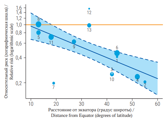
Meta-analysis combines the results of several scientific studies to obtain a summary quantitative estimation of effect size in order to compare the results of several studies and to identify patterns among them and possible sources of biases. Since the research teams, patients, research protocol, clinical guidelines and time intervals are often different between original scientific studies all these sources of difference can influence the results of each study, causing statistical heterogeneity. Meta-analyses are the highest level of credibility within evidence-based medicine, as they allow us to take into account the influence of many confounding factors and publication biases on the true effect size. Understanding the possible sources of erroneous conclusions in papers will allow researchers to analyze the results of such studies correctly and properly plan their own experiments. In this article the reader will be introduced to methods for identifying and quantifying hidden heterogeneity, such as subgroup analysis and meta-regression. In addition, the reader will learn how to calculate effect size in studies, estimate publication bias mathematically and graphically, and incorporate this estimate into the overall average effect size estimate in meta-analyses.
CELL BIOLOGY, CYTOLOGY, HISTOLOGY
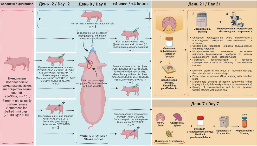
Aim. To study the effectiveness of preventive gene therapy (within 2 days) and gene therapy in the acute phase (after 4 hours) of ischemic stroke in mini-pigs using an autologous leucoconcentrate (AutoLeuc) enriched with recombinant genes of vascular endothelial growth factor (VEGF165), glial cell line-derived neurotrophic factor (GDNF) and neural cell adhesion molecule 1 (NCAM1), as well as the migration of leukocytes transduced with a chimeric adenoviral vector serotype 5 with fiber 35 serotype (Ad5/F35) and the green fluorescent protein (GFP) genome into immune defense organs.
Materials and methods. The experiment was conducted on 8-month-old Vietnamese lop-bellied mini-pigs (n=16). An ischemic stroke was created by occlusion of the distal branches of the left middle cerebral artery and the right common carotid artery. Genetically modified AutoLeuc was administered preventively intravenously 2 days before or in the acute phase 4 hours after stroke modelling; the control group was injected with 30 ml of saline solution. The morphology of the cerebral cortex was assessed using histological methods in the areas bordering the infarction and peri-infarction after 21 days. The migration of genetically modified Ad5/F35-GFP leukocytes into the brain, spleen, and submandibular lymph nodes was studied a week after stroke modelling.
Results. In the peri-infarction zone, the content of pyknotic neurons in control animals was higher, while the number of capillaries was lower than in the gene therapy groups. In the latter, neurons had a typical morphology with preserved outgrowths; in the control group, the outgrowths were tortuous and fragmented. Fluorescence microscopy after injection of AutoLeuc with Ad5/F35-GFP revealed GFP-positive cells in the spleen and submandibular lymph nodes.
Conclusion. 21 days after modeling a stroke in mini-pigs against the background of preventive gene therapy or gene therapy in the acute phase using VEGF165/GDNF/NCAM1 AutoLeuc, greater preservation of neurons and a higher density of capillaries in the peri-infarction zone of ischemic brain damage were established. Leukocytes with Ad5/ F35-GFP were found in the spleen and submandibular lymph nodes.
PATHOLOGICAL PHYSIOLOGY
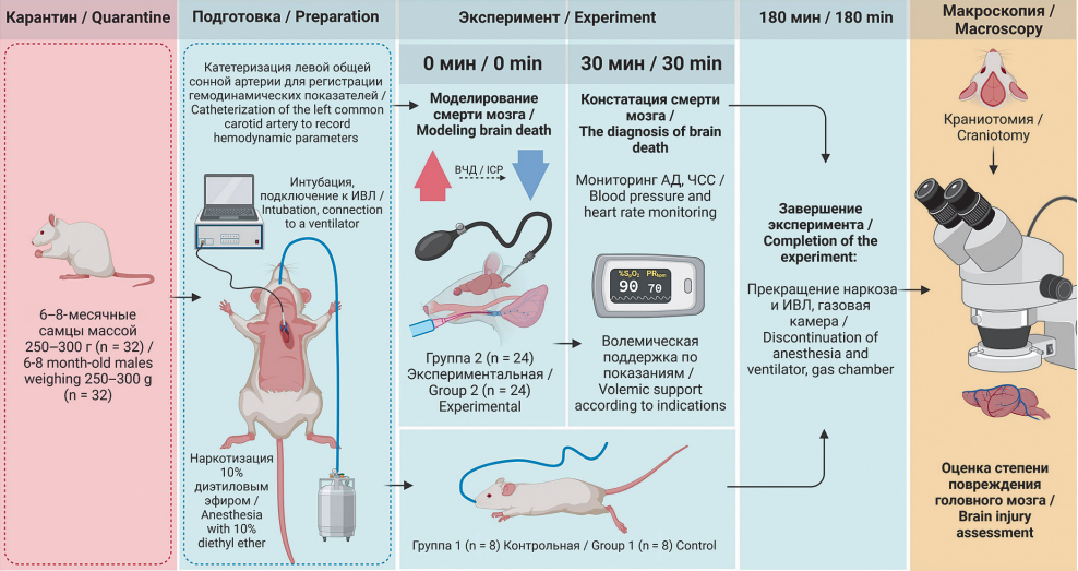
Aim. To test in experiment a pathogenetically adequate model of brain death due to increased intracranial pressure with gradual induction, allowing the evaluation of the changes occurring in the organs of a potential donor.
Materials and methods. 6–8 months old outbred male rats of the experimental group (n = 18) and the control group (n = 8) were anesthetized, the left common carotid artery was catheterized to record systolic, diastolic blood pressure (BP) and heart rate (HR), the mean BP (MBP) was calculated. After transfer to artifi cial ventilation, brain death was simulated in the experimental group using the developed method.
Results. All animals in the experimental group suffered brain death 30 minutes from the start of the experiment;10 rats (56%) died within 3 hours due to progression of circulatory failure. Initially, in anesthetized animals, MBP was 101 (90; 105) mm Hg, HR 310 (297; 315) beats/min. After 5 minutes from the start of brain death induction, MBP increased to 147 (140; 150) mm Hg (p = 0.01), HR to 396 (384; 406) beats/min (p = 0.03). Further, within 20 minutes there was a decrease in MBP to 94 (90; 100) mm Hg and HR to 290 beats/min. During the observation period from 26 to 90 minutes, there was a stabilization of MBP at the level of 87–92 mm Hg, there was a tendency to bradycardia with HR from 263 to 274 beats/min (p = 0.01). Then after 120–150 minutes from the beginning of brain death induction, MBP continued to decrease to 75–80 mmHg (p = 0,03), HR to 256–264 beats/min (p = 0,01). At the end of the experiment, despite volemic support, MBP decreased to 64 (61; 67) mm Hg (p = 0.02), bradycardia worsened with HR to 250 (248; 260) beats/min (p = 0.01), indicating the hemodynamic decompensation.
Conclusion. The results of experimental testing of an animal brain death model on outbred rats showed that this model is pathogenetically adequate and useful to assess the condition of potential donor organs within 3 hours after the induction of brain death.
ONCOLOGY
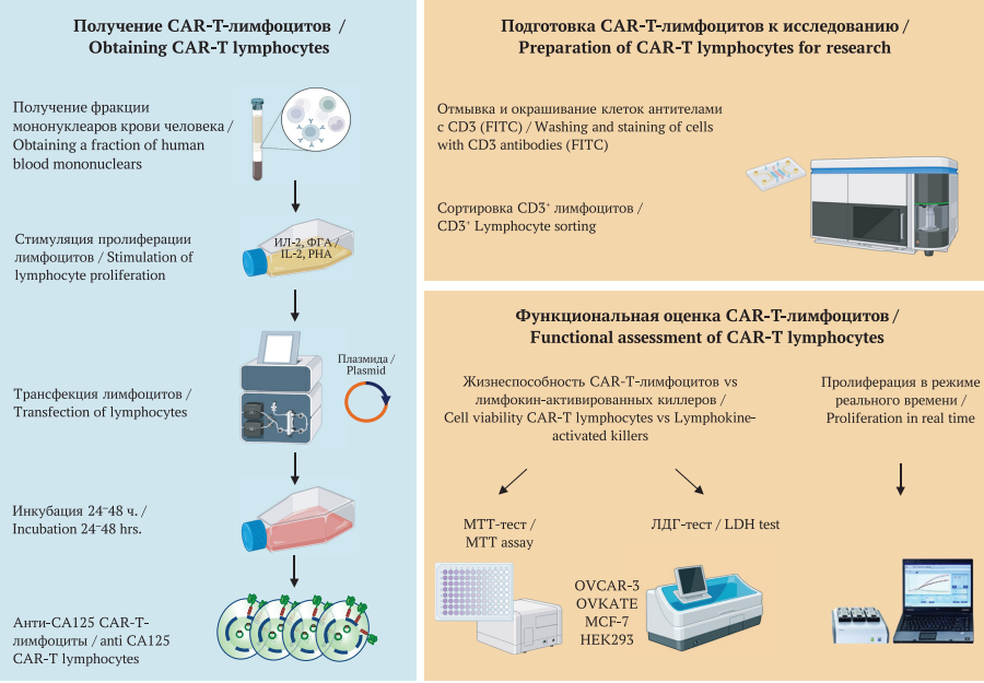
Aim. To evaluate the antitumor efficacy of our developed drug based on cytotoxic T lymphocytes genetically modified with a chimeric antigen receptor (CAR) specific to the CA125 antigen in relation to both CA125-positive and CA125negative cell cultures.
Materials and methods. We performed an in vitro study on CA125-positive human ovarian cancer cells (OVCAR-3, OVKATE) and CA125 negative cells (breast cancer MCF 7, embryonic kidney HEK293). Cytotoxic effects on tumor cells were evaluated after 0, 4, 8 and 24 hours using the 3’-(4,5-Dimethylthiazol-2-yl)-2,5-diphenyl tetrazolium bromide (MTT) and Lactate Dehydrogenase (LDH) tests. We also studied the changes in the number of cells “in real time” when exposed to transfected lymphocytes using the RTCA iCELLIgence device (ACEA Biosciences, USA). Lymphokineactivated killer (LAK) cells were used as a specificity control.
Results. The study demonstrated that anti-CA125 CAR-T lymphocytes exhibited a pronounced cytotoxic effect on OVCAR-3 and OVKATE cell cultures, exceeding the effect of LAK by 1.3 times. The cell population in the experimental samples decreased by 70 ± 4%, which exceeded the LAK effect by 9 ± 8.2%. With regard to the MCF-7 cell line, the cytotoxic effect of anti-CA125 CAR-T lymphocytes was minimal as evidenced by a 25.8% decrease in the relative number of live cells in comparison to the LAK cytotoxicity of 68%. Real-time monitoring of cell proliferation and viability proved a high specific cytotoxic effect of anti-CA125 CAR-T lymphocytes against tumor cultures expressing CA-125, while inferior to LAK in cultures not expressing CA125 (MCF-7, HEK293).
Conclusions. The use of anti-CA125 CAR-T lymphocytes against CA125-positive tumor cell lines OVCAR-3 and OVKATE demonstrated a pronounced specific cytotoxic effect exceeding the cytotoxic effect of LAK, which was not achieved against CA125-negative MCF-7 and HEK293 cells.
INTERNAL MEDICINE
Aim. To study the characteristics of the fatty acid (FA) profi le of blood serum and erythrocyte membranes in patients with two forms of fatty liver disease (metabolic + alcoholic): steatosis and steatohepatitis with normal transaminase activity.
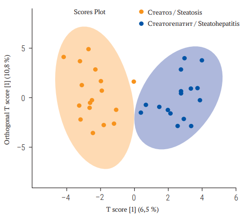
Materials and methods. We examined 33 men (50.7 ± 9.6 years) with fatty liver disease (metabolic and alcoholic) with fi brosis F ≤ 1 (FibroTest). According to the ActiTest results, patients were divided into groups of steatosis – with minimal (A0–1) activity (n = 17) and steatohepatitis – with moderate/severe (A2–3) necroinfl ammatory activity (n = 16). The FA composition of blood serum and erythrocyte membranes was studied using gas chromatography/mass spectrometry Agilent 7000B (Agilent Technologies, Inc., USA). Methods of unpaired statistics using volcano plot and discriminant analysis based on orthogonal least squares (Orthogonal Partial Least Squares Discriminant Analysis, OPLS-DA), ROC analysis were applied.
Results. Volcano plot analysis showed that in patients with fatty liver disease (metabolic and alcoholic) with normal transaminase activity, serum levels of stearic C18:0 (p = 0.016), arachidic C20:0 (p = 0.023), ratio saturated / polyunsaturated fatty acids (PUFA) (p = 0.001) were statistically signifi cantly higher in the steatohepatitis group compared with the steatosis group. The total content in the blood serum of all PUFA (p = 0.003), margaric C17:0 (p = 0.011), the sum of two omega-3 PUFA – eicosapentaenoic acid (C20:5n-3) and docosahexaenoic acid (C22:6n-3) (p = 0.04), the total content of all omega-3 PUFA (p = 0.042) were statistically signifi cantly lower in patients with steatohepatitis. OPLS-DA demonstrated fairly accurate separation of steatohepatitis and steatosis using individual FA and their ratios. When individual FA and their ratios were included in the analysis, a model was obtained with AUC = 0.827 (95% confi dence interval 0.499–1.0), sensitivity 82.2% and specifi city 80.7%.
Conclusion. FA in blood serum and erythrocyte membranes appear to be promising biomarkers of steatohepatitis with normal levels of transaminases.

ISSN 2658-3348 (Online)


