


EDITORIAL


CELL BIOLOGY, CYTOLOGY, HISTOLOGY
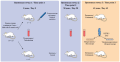
The ability of mesenchymal stromal cells (MSCs) to migrate into tissue defects and stimulate regeneration makes them a valuable resource for cell therapy. However, in many cases, in vitro cultivation and the influence of the pathological microenvironment in the patient’s body reduce the viability and therapeutic efficacy of MSCs, so their regenerative potential needs to be strengthened. Preconditioning with hormones, cytokines, various chemical or physical factors, cultivation in a three-dimensional environment or at a reduced oxygen content improves the ability of MSCs to colonize damaged tissue, survive in it, and produce regulatory molecules for regeneration. The same goals can be achieved by genetic modification of MSCs. In addition, with the help of transfected MSCs, it is possible to deliver genes necessary for the treatment of hereditary or oncological diseases into the tissue. Finally, an alternative to avoid a decrease in the therapeutic potential of subsequently transplanted MSCs during cultivation can be stimulation of the migration of endogenous patient cells from tissue niches through the systemic circulation to the area of damage. The development of these approaches opens the way to increasing the efficiency of using MSCs in regenerative medicine.
Aim. To determine the delayed (after 2 months) effect of spinal cord injury (SCI) in the lower thoracic region in the mini-pigs on the morphologic state of macro- and microglia in nearby and remote caudal areas.
Materials and methods. Sexually mature female Vietnamese pot-bellied pigs were randomly divided into two groups: SCI (n = 3) and intact (n = 3). Dosed contusion SCI was modelled at the level of the Th8–Th9 vertebrae, and transverse cryostat sections of the caudal segment adjacent to the epicenter of injury and the lumbar thickening (L4–S2) were examined 2 months later. The expression of astrocyte markers (glial fibrillary acidic protein, GFAP) and microglial markers (ionized calcium-binding adapter molecule 1, Iba1) was assessed as the relative immunopositive area occupied by cells. When counting the number of oligodendroglial cells (oligodendrocyte transcription factor 2, Olig2), the presence of nuclei detectable with 4’,6-diamidino-2-phenylindole (DAPI) was taken into account.
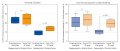
Results. After SCI, an increase in the relative areas occupied by GFAP-positive astrocytes and Iba1-positive microglia and a decrease in Olig2-positive oligodendrocytes were detected in both the lesion area and lumbar thickening. In both regions, 2 months after SCI, the proportion of astrocytes was not significantly different in the anterior horns and doubled in the posterior horns. Microglia cells with SCI were 2.5 times more in the anterior horns of both regions and in the posterior horns of the lumbar thickening, while the presence of microglia increased slightly (1.2 times) in the posterior horns in the SCI region. The number of oligodendrocytes decreased in the area of the epicenter of SCI in the anterior and posterior horns by 1.5–1.75 times, and in the lumbar thickening more significantly: the number decreased by 2.5 times in the anterior horn and 5.5 times in the posterior horn.
Conclusion. The results of the study revealed a similar pattern of macro- and microglial cell distribution both in the SCI region and in remote areas. The obtained data testify to the necessity to take into account the state of the areas of nervous tissue remote from the epicenter of SCI when stimulating neuroregeneration in such patients
Aim. We aimed to determine the content of neurons expressing somatostatin (SST) and their colocalization with cells expressing tyrosine hydroxylase (TH) and neuropeptide Y (NPY) in the cranial cervical ganglion (CCG) and celiac plexus in rats.
Material and methods. We used 30 white male Wistar rats of six age groups (5 rats per group): newborn pups, 10-, 20-, 30-, and 60-day-old pups, and 24-month-old pups. We incubated their ganglia sections with primary antibodies against SST, NPY, and TH, as well as with secondary antibodies conjugated with fluorochromes. We evaluated the ratio between immunoreactive (IR) neurons with a visible nucleolus and excessive fluorescence and the total number of neurons, as well as the average cross-sectional area, by ImageJ software (NIH, USA).
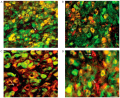
Results. SST-IR neurons were not found in the CCG. However, the immunoreaction (as granules) was revealed in most perikaryons at the celiac plexus for SST and NPY with a rather homogeneous distribution for TH. The ratio of ST-IR neurons reached 33% in pups, doubled during the first month of life, and then remained constant (70–73%). No statistically significant differences were found between the ratios of SST-IR neurons of the cranial mesenteric ganglion (CMG) and celiac ganglion (CG) for all age groups. From the moment of birth to 60 days of life, the average cross-sectional area of SST-IR neurons in the CG and CMG increased by 3.4–3.9 times and then did not change until 24 months. From the 20th day of life, the average cross-sectional area of SST-IR neurons in the CG was significantly higher than that in the CMG. All SST-IR neurons in all age groups expressed TH, while 90–94% of neurons expressed NPY.
Conclusions. The content of ST-IR neurons in different sympathetic nodes is not the same: they are absent in the CCG, and their ratio and area in the celiac plexus increase during early postnatal development. This may be due to the peculiarities of innervated target organs.
Aim. To study the morphology of neurons in the cerebral cortex of rat pups on day 20 under conditions of administration of a nitric oxide synthase inhibitor (NOS) during placentation.
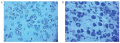
Materials and methods. Outbred white female rats (n = 12) were randomly divided into 2 groups of 6 rats each. On the 11th day of pregnancy, the experimental group received a single intramuscular injection of N(omega)-nitro-L-arginine methyl ester (L NAME) at a dose of 25 mg/kg, in the control group – once intramuscularly 0.9% NaCl solution. Born rat pups were randomly selected one from the mother. On the 20th day, after medical euthanasia, the brain was collected. In the anterior part of the frontal cortex, we studied the density and area of neurons, the size and shape of perikarya and the severity of their staining with toluidine blue.
Results. In the experimental group of 20-day-old rat pups, compared to the control group, the density and area of neurons were less by 10% (p > 0.05) and 22% (p > 0.05), respectively, the shape of the perikarya also changed to elongated, the elongation factor increased by 0.3 units. (p < 0.05) and there was a sixfold increase in the proportion of hyperchromic neurons (p < 0.05), hyperchromic wrinkled (p < 0.001) neurons appeared, which were absent in control animals.
Conclusion. Morphological changes in neurons of the cerebral cortex in rat pups born from females who received a NOS inhibitor during placentation may be a consequence of a decrease in the formation of nitric oxide in the neurons themselves and in the endothelium of the vessels supplying the brain
Aim. We aimed to study the histological and ultramicroscopic structure of the striated muscle tissue of the external anal sphincter (EAS) of mature male rats under experimental androgen deficiency.
Materials and methods. The study included 10 male laboratory rats aged 8 months, which were randomly divided into 2 groups of 5 each. The experimental group underwent bilateral orchiectomy to create testosterone deficiency. After 45 days, rats were sacrificed. We studied the concentration of testosterone in histological sections of EASs using light microscopy and ultramicroscopy. We also determined the diameter of muscle fibers and the thickness of endomysium, the area of muscle fibers, connective tissue, myofibrils and cytoplasm, identification of glycogen granules in the cytoplasm and intermyofibrillar space, as well as changes in mitochondria.
Results. In the experimental group, on the 45th day after surgical castration, the testosterone level was 2.5 times lower than in the control group 2.69 (2.52; 2.73) nmol/l vs. 7.20 (6.83; 7.21) nmol/l, p = 0.008. Using morphometric analysis, we found that in the experimental group after surgical castration the diameter of the muscle fibers was statistically significantly smaller than in the control group: 6.56 (3.96; 7.24) µm vs. 9.52 (8.88; 10.44) µm, p < 0.001, while the thickness of the endomysium in the experimental group was greater: 3.34 (3.11; 3.78) µm vs. 1.62 (1.51; 1.86) µm, p < 0.0001. The ratio of muscle fiber area/connective tissue area was statistically significantly lower in the group after castration: 1.64 (1.50; 1.78) vs. 4.00 (3.17; 5.25), p < 0.0001. The ratio of myofibril area/cytoplasmic area changed in the experimental group towards the predominance of cytoplasm 0.79 (0.67; 0.79) vs. 5.25 (5.25; 7.33), p < 0.0001. With an increase in cytoplasmic volume, an increase in the number of glycogen granules was observed; pathological forms of mitochondria were identified: swelling, destruction of cristae and vacuolization of their matrix.
Conclusion. Under conditions of testosterone deficiency, along with atrophic processes, compensatory and adaptive mechanisms are formed in the striated skeletal muscle tissue of the EAS, aimed at restoring its metabolic and functional organization

ISSN 2658-3348 (Online)


