


MODELING IN MEDICINE
Diagnostic efficiency of breast cancer screening remains one of the most important issues in oncology and radiology. Artificial intelligence technologies are widely used in clinical medicine to effectively solve a number of technological and diagnostic problems.
The aim. To develop segmentation neural network model for breast plain radiographs analysis with subsequent study of its clinical effectiveness.
Materials and methods. The artificial intelligence-based system was developed to analyze X-ray mammography, аnd included a segmentation neural network with the U-Net architecture, a classification neural architecture ResNet50 with outputting the result using gradient boosting. 15486 X-ray cases were used for training, estimation of diagnostic accuracy and validation of the developed segmental model. All cases were labeled in specially developed software environment LabelCMAITech. The segmentation accuracy was determined by Intersection over Union (IoU) similarity coefficient, the probability of malignancy was calculated using the binary classification metrics.
Results. The developed system is represented by a segmentation model based on neural network architecture. The model allows, with high accuracy of 0.8176 and higher, at threshold values on the output neurons of the network of 0.1 and 0.15, to localize X-ray findings that have diagnostic value for determining the likelihood of the presence of breast cancer signs in an X-ray mammographic study — focus, architecture distortion, local asymmetry, calcifications. When comparing the results of machine segmentation and marking of images by a radiologist, it was found that the model is not inferior to the doctor in the accuracy of determining the formations, extra-focal calcifications and intraglandular lymph nodes.
Conclusion. The results of this study allow considering the model as an intelligent assistant to a radiologist in the analysis of screening mammographic cases.
SURGERY
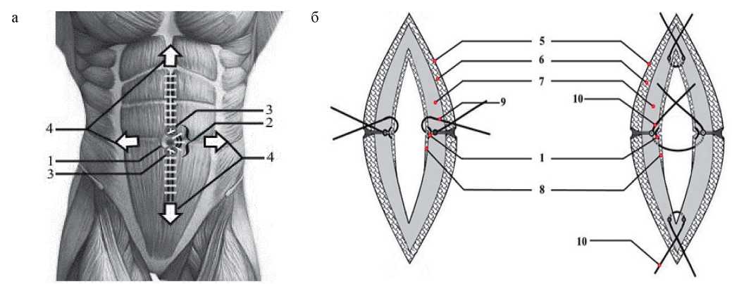
The problem of postoperative ventral hernias remains relevant due to the high frequency of their development — in 7–24% of patients.
Aim. To evaluate the effectiveness of prevention of postoperative ventral hernias using a new technique of access to the abdominal cavity in comparison with traditional laparotomy.
Materials and methods. The proposed new technique of access to the abdominal cavity along the midline of the abdomen with dissection of the navel and umbilical ring was carried out in accordance with the method developed by us, registered by the patent of the Russian Federation. For the final analysis, 134 patients were selected, divided into group 1 (n = 67), in which the median access was performed using a new technique, and group 2, in which the traditional median access was performed with the left umbilical ring bypass (n = 67). The initial parameters of patients, characteristics during and after surgery were evaluated. The duration of follow-up after surgery was 24 months.
Results. Groups 1 and 2 were comparable by gender, age, body mass index, and the presence of comorbidities. In 79% of patients in group 1 and in 67% of patients in group 2 (the difference is not significant), indications for surgery were malignancies of the abdominal cavity. The groups did not differ in the types of median laparotomy, the time of surgery, the amount of blood loss, the time of removal of postoperative sutures, and the duration of hospitalization. All patients were followed up for 24 months. Postoperative ventral hernia developed in one patient (1.5%) in group 1 and in 5 (7.5%) patients in group 2 within 12 to 24 months after surgery. There were no statistically significant differences in the frequency of hernia development (Mantel — Cox test, p = 0.100)
Conclusion. A new method of median laparotomy in the treatment of patients with planned surgical pathology of the abdominal cavity is characterized by a low rate of postoperative hernia development.
INTERNAL MEDICINE

The presence of arterial hypertension (AH) leads to the development of cognitive dysfunction, in the genesis of which a significant role is assigned to vascular factors.
Aim. To study the state of cognitive function and associated vascular factors in patients with uncontrolled AH.
Materials and methods. The research involved 88 patients with uncontrolled AH (UAH) — group 1 (median age 60, men — 39%) and 46 patients with controlled AH (CAH) — group 2 (median age 59, men — 41%). Cognitive function was assessed using the Montreal Cognitive Assessment (MoCA). There were studied vascular factors: thickness of the intima-media complex (IMC), pulse wave velocity (PWV), microcirculation flow index (MFI) and asymmetric dimethylarginine (ADMA) concentrations. For the statistical analysis the following criteria were used: Student t-test, Mann—Whitney test. Multifactorial linear regression analysis was performed in groups.
Results. In Group 1, there was a lower cognitive function index by MoCA — 24 [22; 26] points against 26 [25; 27] points in Group 2 (p = 0.002). IMC thickness was higher in Group 1 than in Group 2 (1.1 [0.90; 1.20] mm vs 1.0 [0.80; 1.10] mm, p = 0.042), concentration of ADMA was higher in Group 1 (0.73 ± 0.21 µmol/l vs 0.65 ± 0.1 µmol/l, p = 0.02), MFI was higher in Group 2 (30.6 [27.1; 34.4] perf. units vs. 22.8 [18.6; 26.1] perf. units, р < 0.001). No differences between the groups were found in PWV. In regression analysis, the following factors had a statistically significant effect on MoCA scores: in Group 1 — age, IMC thickness, ADMA and MFI; in Group 2 — age and glomerular filtrate rate.
Conclusion. Patients with uncontrolled AH have more pronounced cognitive dysfunction than those with controlled AH, which is associated with increased IMC thickness, impaired microcirculation and increased ADMA concentration.
Hereditary predisposition is the most significant risk factor for Crohn’s disease (CD) in siblings.
Case reports. CD was diagnosed in 3 brothers in a family with 6 children of the same generation: the disease manifested itself in one — at the age of 15 years and in two brothers — at the age of 17 years. In the older brother, CD manifested with acute intestinal obstruction, ileum perforation, diffuse peritonitis; in the middle — with symptoms similar to appendicular infiltrate. Both brothers underwent resection of the terminal ileum, cecum, and part of the ascending colon with the imposition of ileoascendoanastomosis. The younger brother was diagnosed with a non-stricturing, non-penetrating form of CD with a gradual onset and no complications. Among the siblings without CD, two have rheumatoid arthritis and vitiligo.
Discussion. Similar signs of the familial form of CD were young age of manifestation and ileocolonic disease location; distinctive signs — the presence of complications and surgical interventions in only two brothers. A special feature of the case reports is the CD development only in boys and the combination of three different autoimmune diseases in one generation of the family.
ОBSTETRICS AND GYNECOLOGY
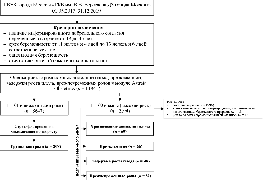
Effect of maternal factors on indicators of increased risk of chromosomal abnormalities (CA), pre-eclampsia (PE), Small-forGestational-Age Fetus (SGA fetus) and preterm labour and birth (PB) during prenatal screening has not been sufficiently studied.
Aim. To study the effect of maternal reproductive factors on the risk indicators of CA, PE, SGA fetus and PB, assessed during prenatal screening using the Astraia Obstetrics module.
Materials and methods. Of the 11,841 pregnant women who were prenatal screened, 18.53% of the patients had at high risk of the outcomes studied (frequency 1: 100 and above). The subgroup of isolated high risk for CA included 69, PE — 66, SGA fetus — 48, PB — 52 patients. From the group of patients with low risk, 208 patients were selected for the control group by the method of stratified randomization by age.
Results. Among extragenital diseases, the most common in all high-risk subgroups were: hypertension (AH) I and II degree — 31–47% versus 4.8% of the control group (p < 0.05), varicose veins of the lower extremities (VVLE) — 17–30% vs. 5.3% in the control group (p < 0.05), a history of ovarian tumor — 12–33% vs. 3% in the control group (p < 0.05). In the high-risk subgroups for the development of CA, PE and SGA fetus, fibroids uterus and iron deficiency anaemia (IDA) were more common compared to control: 10–41% vs. 1% (p < 0.05) and 10–17% vs. 3% (p < 0.05), respectively (p < 0.05). Primiparas with a history of pregnancy were more common in subgroups with a high risk of CA (33%) and PR (35%) versus 17% in controls.
Conclusion. An association has been established between high risk for all the outcomes studied and AC, VVLE, history of ovarian tumor. High-risk subgroups for CA, PE and SGA fetus have a higher incidence of uterine fibroids and IDA compared to control.
PATHOLOGICAL PHYSIOLOGY
Lymphangiogenesis plays an important role in development of renal parenchyma inflammation during kidney injury. Vascular endothelial growth factor type C (VEGF-C), cytokine that regulates lymphangiogenesis, is a potential early biomarker for acute kidney injury.
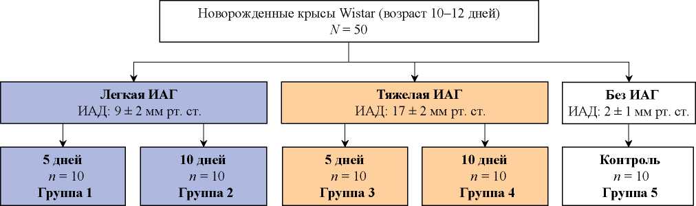
Aim. To study the concentration of VEGF-C in renal homogenate and blood serum of newborn rats with experimental intraabdominal hypertension (IAH) of varying severity and duration, to establish a relationship with morphological changes in the renal tissue.
Materials and methods. The experiment was conducted on 50 newborn Wistar rats. Rats were divided into 5 groups of 10 rats each: groups 1 and 2 with mild IAH lasting 5 and 10 days, respectively, and groups 3 and 4 with severe IAH lasting 5 and 10 days, respectively, and the control group. IAH was modelled by injecting sterile vaseline into the abdominal cavity to a predetermined level of IAH under the control of intra-vesical manometry. VEGF-C content was measured by ELISA. Morphological examination of the biopsy material and its photography were carried out using a Leica DM2000 microscope. The Mann—Whitney, Kruskal—Wallis, Wilcoxon tests, as well as one-way ANOVA, were used for statistical analysis.
Results. The level of VEGF-C in the renal homogenate was increased in all groups (pc < 0.001); the degree of VEGF-C increase depended on the severity of IAH (p < 0.05) but not on the duration of IAH exposure. The VEGF-C blood serum level was increased only in group 3 (pc = 0.011). Morphological analysis showed hydropic dystrophy: changes in the height of the tubular epithelium, an increase in interstitial edema, expansion of the urinary spaces of glomeruli. The degree of morphological changes depended on the severity and duration of IAH.
Conclusion. Changes in VEGF-C level assessed in the renal homogenate correlated with morphological changes in renal tissue of rats with different severity and duration of IAH.
CELL BIOLOGY, CYTOLOGY, HISTOLOGY
Kidney diseases are an important medical problem. Kidney injuries are accompanied by oxidative stress, cell death, capillary destruction, inflammation and fibrosis. Mesenchymal stromal cells (MSCs) have a complex effect on the regeneration by producing various regulatory molecules, including those inside extracellular vesicles, and therefore are considered as a promising therapeutic resource for cell therapy of kidney diseases. Their renoprotective effect has been shown in different experimental models, but the results of the clinical trials are ambiguous. Clinical use of MSCs is complicated by their low survival rate in the injured kidney, potential immunogenicity, tumorogenicity and fibrogenicity. Cell-free therapy with the secretory products of MSCs such as conditioned environments or extracellular vesicles is a promising direction for using their regenerative potential. However, introduction of MSCs and their secretory products into medical practice requires further research into the mechanisms of their proregenerative action, improvement of cultivation protocols, and more clinical trials.
RETRACTION

ISSN 2658-3348 (Online)


